- General Dermatology
- Eczema
- Alopecia
- Aesthetics
- Vitiligo
- COVID-19
- Actinic Keratosis
- Precision Medicine and Biologics
- Rare Disease
- Wound Care
- Rosacea
- Psoriasis
- Psoriatic Arthritis
- Atopic Dermatitis
- Melasma
- NP and PA
- Skin Cancer
- Hidradenitis Suppurativa
- Drug Watch
- Pigmentary Disorders
- Acne
- Pediatric Dermatology
- Practice Management
Diagnosing, treating vesiculobullous dermatoses
In this article, Joseph C English III, M.D., professor of dermatology at the University of Pittsburgh, offers his insight for diagnosing and treating cutaneous blistering diseases. Participate in this forum.


“You have to be able to distinguish clinically and sometimes histologically what’s going on in order to make the correct diagnosis,” says Dr. English, who presented on vesiculo-bullous dermatoses at the Society of Dermatology Physician Assistants (SDPA) annual fall conference in October 2018. (Photo courtesy of Joseph C English III, M.D.)
Diagnosing cutaneous blistering diseases can be simple to complex, according to Joseph C English III, M.D., professor of dermatology at the University of Pittsburgh.
“You have to be able to distinguish clinically and sometimes histologically what’s going on in order to make the correct diagnosis,” says Dr. English, who presented on vesiculo-bullous dermatoses at the Society of Dermatology Physician Assistants (SDPA) annual fall conference in October 2018.
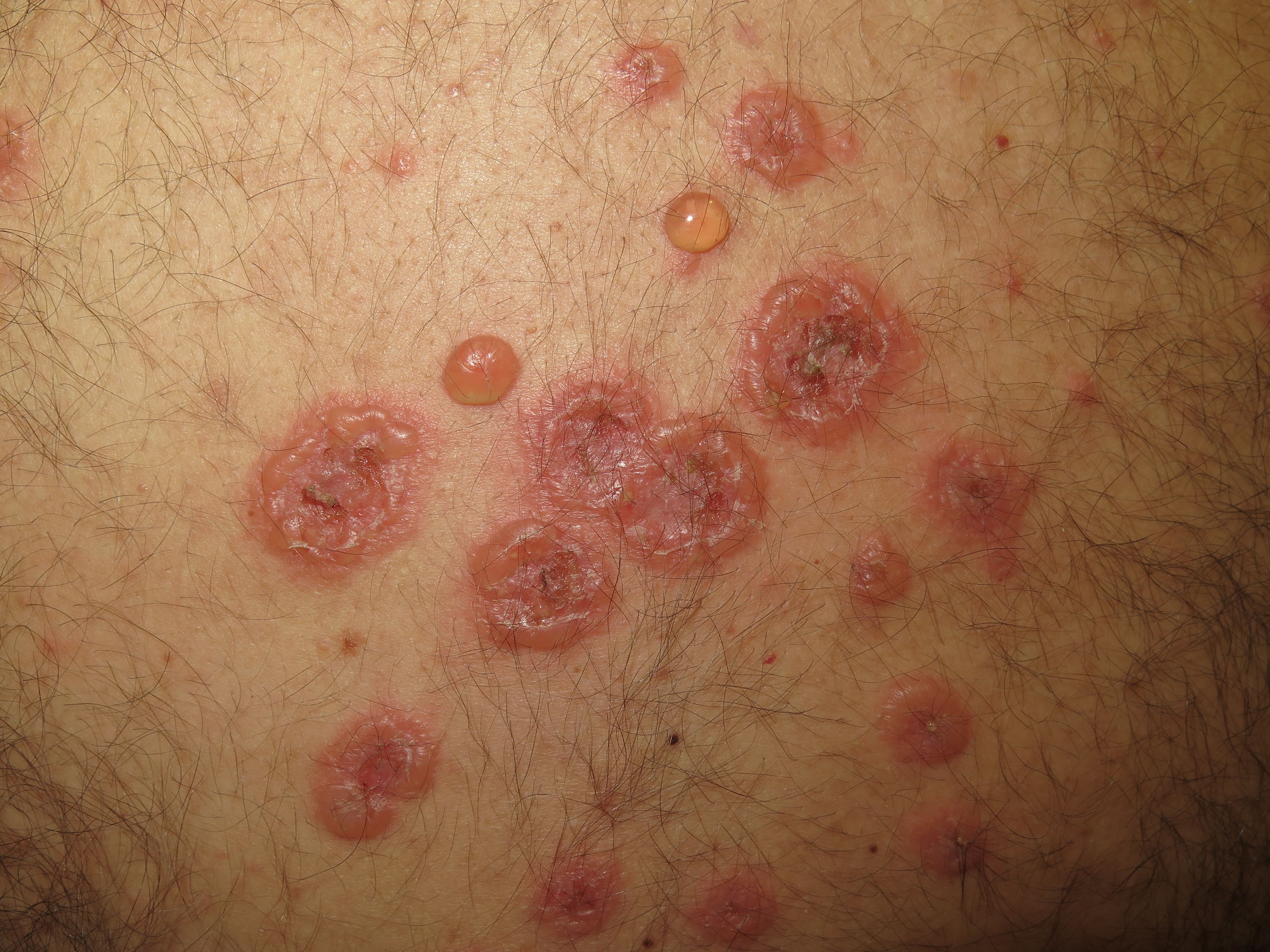
Vesicles are fluid-filled, or serosanguinous, skin lesions that are 1 to 10 mm in size, while bullae are larger at more than 10mm. Often these blisters present as a combination on the skin, known as vesiculo-bullous dermatoses, according to Dr. English.
MAKING A DIAGNOSIS
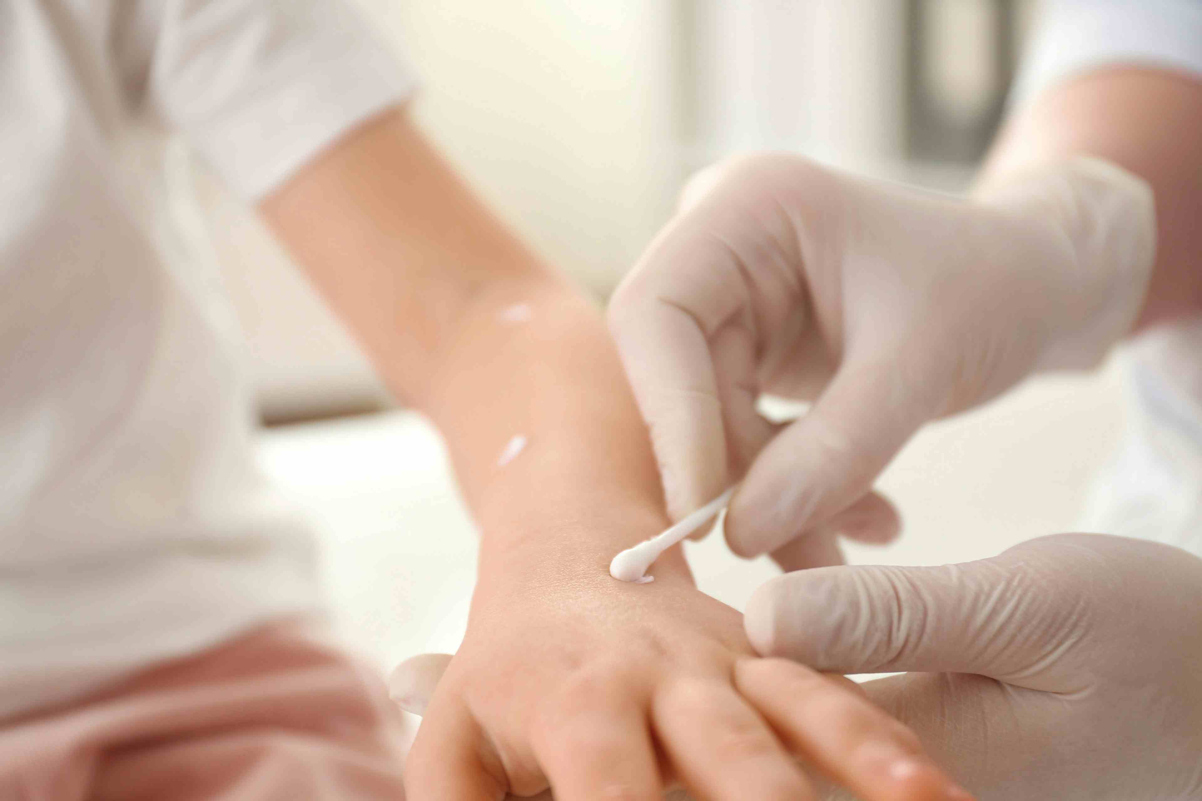
There are a lot of things one can do to help determine the diagnosis of vesicular disorders, including culturing fluid from the vesicle for bacteria, according to Dr. English.
Other options include in-office microscopy; the Tzanck preparation, to test for herpes infections; potassium hydroxide, KOH, preparation to check for fungal infections; or ectoparasite preparation to detect a scabies infestation.
“When the lesions are more widespread, performing a cutaneous biopsy for histopathology and direct immunofluorescence to determine if it’s an autoimmune blistering disorder or other process is required,” he says.
MAKING A DIFFERENTIAL DIAGNOSIS
Providers looking at these lesions in the clinic should take note of whether they’re localized or generalized eruptions. There are several possibilities when it comes to blistering.
“Limited vesiculo-bullous diseases commonly seen in clinic are allergic or irritant contact dermatoses or arthropod assaults. Lower leg bullous eruptions can be from lymphedema or diabetic bullae in poorly controlled diabetic patients,” Dr. English says. “You can see vesicles in the setting of trauma or pressure. Sometimes blisters are not the primary process but rather secondary from cutaneous leukocytoclastic vasculitis. Certain infections can be bullous, including bacterial, fungal and viral, especially herpes infections.”
Most concerning are generalized vesiculo-bullous eruptions such as severe cutaneous adverse reactions from medications and autoimmune bullous disorders. Drug eruptions can cause erythema multiforme, Stevens-Johnson Syndrome (SJS) and toxic epidermal necrolysis. While pemphigus vulgaris, paraneoplastic pemphigus, bullous pemphigoid and linear IgA bullous disease are examples of widespread immunobullous disease.
MANAGING CUTANEOUS BLISTERING
“Once you get the diagnosis, you can determine more specific treatment,” according to Dr. English.
Examples of treatment are topical high potency steroids for an inflammatory arthropod reactions and oral antiviral treatment (valacyclovir) for viral eruptions, such as herpes simplex virus or herpes zoster.
“In cases where it’s a drug reaction, it depends on the extent and nature. If it’s a minor eruption you can usually treat it topically. Sometimes adding an oral steroid taper may be required,” he says. “But if it’s a more severe cutaneous adverse reaction, such as Stevens-Johnson Syndrome or Toxic Epidermal Necrolysis (TEN), hospitalization, discontinuance of offending medication and the use of intravenous immunoglobulin (IVIG), cyclosporin or even the biologic etanercept can be beneficial in decreasing morbidity and mortality.1, 2
“It’s important to have good wound care or supportive care in a burn unit, if possible, to treat severe blistering cases,” he says.
Therapy for the pemphigoid or pemphigus group disorders usually starts with prednisone and if needed nonsteroidal anti-inflammatory agents, such as mycophenolate mofetil, rituximab and IVIG, can be used according to Dr. English.
“Trying to find anti-inflammatory regimens for the autoimmune blistering disorders is often a complicated process to determine the quickest and safest way to improve those diseases,” he says.


Disclosure: None
References:
1. Kirchhof MG, Miliszewski MA, Sikora S, Papp A, Dutz JP. Retrospective review of Stevens-Johnson syndrome/toxic epidermal necrolysis treatment comparing intravenous immunoglobulin with cyclosporine. J Am Acad Dermatol. 2014 Nov;71(5):941-7. doi: 10.1016/j.jaad.2014.07.016. https://www.jaad.org/article/S0190-9622(14)01679-X/fulltext.
2. Paradisi A, Abeni D, Bergamo F, Ricci F, Didona D, Didona B. Etanercept therapy for toxic epidermal necrolysis. J Am Acad Dermatol. 2014 Aug;71(2):278-83. doi: 10.1016/j.jaad.2014.04.044.