- General Dermatology
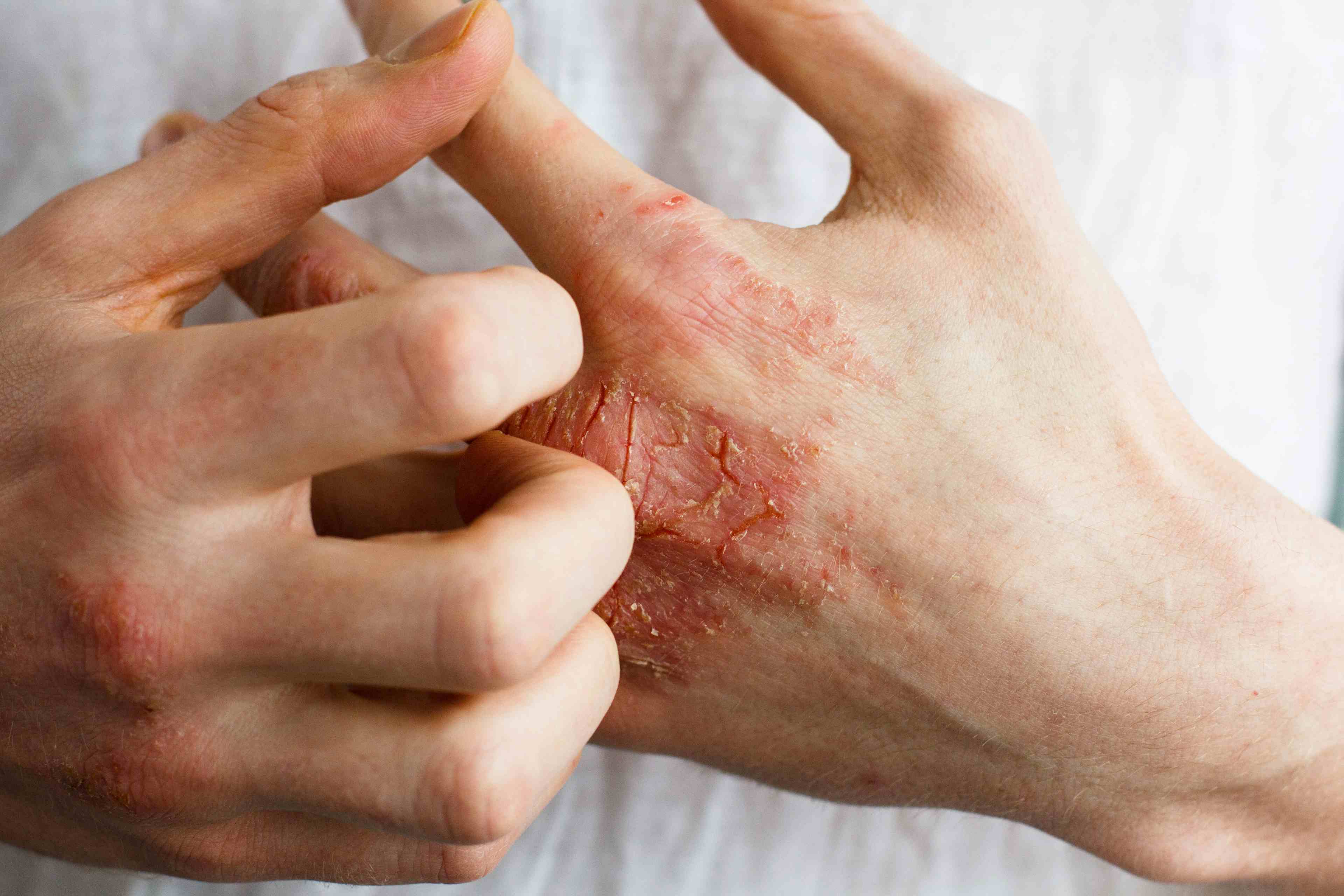
- Eczema
- Alopecia
- Aesthetics
- Vitiligo
- COVID-19
- Actinic Keratosis
- Precision Medicine and Biologics
- Rare Disease
- Wound Care
- Rosacea
- Psoriasis
- Psoriatic Arthritis
- Atopic Dermatitis
- Melasma
- NP and PA
- Skin Cancer
- Hidradenitis Suppurativa
- Drug Watch
- Pigmentary Disorders
- Acne
- Pediatric Dermatology
- Practice Management
Lipid Imaging Mass Spectrometry Aids in Accurate Skin Cancer Diagnosis, Study Says
Investigators used LIMS to classify melanoma samples as tumors and non-tumors.


Yulia/Adobe Stock
For melanoma, the most significant factors that aid patient survival are early detection and precise diagnosis. Lipid imaging mass spectrometry (LIMS) can help achieve accurate diagnoses by directly enabling the exploration of tissue sections by scanning the samples. In a study published in International Journal of Cancer, investigators used LIMS to classify melanoma samples as tumor and non-tumor, as well as identify whether the samples are metastatic or primary tumors.
The 53 enrolled patients were aged 19 to 96 years with a similar gender distribution. Samples and data from patients were collected from 2017 to 2020 and provided by 3 different centers.
A total of 15 nevi, 24 primary melanomas, and 14 metastases were collected, in addition to clinical information including patients’ gender, age at primary tumor, localization of primary melanoma, localization of metastasis, stage, and histological subtypes (superficial spreading melanoma [SSM], nodular melanoma [NM], acrolentiginous melanoma [ALM], and lentigo maligna melanoma [LMM]). Most of the nevi were intradermal (n = 11); however, a compound nevus and a junctional nevus were also included. Primary melanoma samples corresponded to SSM (n = 19), NM (n = 2), LMM (n = 1), and ALM (n = 2). Further, all metastasis samples were diagnosed as locoregional in transit metastasis.
Collection was performed by gathering 4 μm-thick sections for hematoxylin-eosin (H&E) staining and for immunohistochemistry (IHC) of Melan A and HMB45 melanocyte biomarkers. Melan A—also called melanoma antigen recognized by T cells (MART-1)—marks both melanocytes from nevi and melanoma, whereas HBM45 specifically marks melanoma cells. Next, 16 μm-thick sections were obtained for LIMS and were selected using H&E and IHC imaging. Due to the speed of the mass spectrometer and lipid propensity to oxidation, the entire area of the sections was unable to be explored. The sections explored by LIMS were stained with H&E allowing for the annotation of histological areas and structures to correlate them with the segments obtained from the analysis of the LIMS images.
Using LIMS, data from each section were analyzed using a segmentation algorithm to isolate and identify the lipid signatures of each histological area in the section. A heuristic approach was used to establish the number of segments on each image, with the initial number of segments being set to 10 for nevus and melanoma, and 5 for metastasis. Segments suggested by the algorithm were verified by examining their correlation, and the segments with correlations higher than 95% were grouped together due to the high correlation indicating similar histologic areas. Each segment was assigned a color based on correlation, and the 2 segments with the lowest correlation were assigned the colors at both ends of the scale with the rest of the segments receiving colors in between.
Melanocytic lipid signatures come from nevus sections, whereas more than 1 lipid fingerprint was extracted from each primary and metastatic melanoma sample. A noticeable separation of melanocytic samples from those of tumor cells was observed during the training set, with incomplete separation being observed between melanoma and metastasis; however, the analysis of the samples in the validation set using only those lipid species showed statistically significant differences in the training set—potential biomarkers—resulting in a perfect separation of the 3 types of samples.
The lipid fingerprints of nevus melanocytes were compared with those of primary melanoma and metastasis, and a perfect separation was achieved in all cases, highlighting the strong metabolic changes that accompany tumor transformation. Further, classification of primary and metastatic samples was not perfect in the training set, with only 2 of the metastasis misclassified as primary tumors; however, sample classification was perfect in the validation group using the potential lipid biomarkers.
The results indicate that a substantial change in the lipid fingerprint accompanies tumor transformation, and these changes enable building classification models with performances between 90% and 100%—algorithm-dependent—when biopsies from nevus, primary melanoma, and metastatic melanoma are included and with perfect performance in binary comparisons. LIMS can be used alongside with classifiers to help identify tumor cells directly from tissue sections. With the correct interpretation, the technique reports important information on the tumor microenvironment, providing valuable insights in prognosis, and can even help with the early and accurate diagnosis of melanoma. This presents opportunities for the use of this technique in clinics for faster and automated sample screening.
Further, lipidomic changes were observed in both primary melanoma and metastasis, with focuses on phospholipids (LPI, LPI-O/P), prostaglandins (PG), and phosphatidylserines (PS). Primary melanoma demonstrates a decrease in certain LPI species, which may indicate a higher demand for arachidonic acid for inflammation-related mediators. Fewer PG and PS species were detected, with a shift toward monounsaturated and polyunsaturated fatty acid-containing species.
Limitations of the study include the small sample size enrolled and the need for larger cohorts to validate the findings. Further, the investigators acknowledge that the study was limited to negative ion mode LIMS, and that other modes of imaging mass spectrometry could help provide additional, in-depth information. The study authors recommend future advancements can include the creation of a library of lipid fingerprints that can automatically annotate tissue sections, as well as integration of this technology into other types of cancer.
Reference
Huergo-Baños, C, Velasco, V, Garate, J, et al. Lipid fingerprint-based histology accurately classifies nevus, primary melanoma, and metastatic melanoma samples. Int J Cancer. 2023; 1-11. doi:10.1002/ijc.34800
[This article was originally published by our sister publication, Pharmacy Times.]