- Acne
- Actinic Keratosis
- Aesthetics
- Alopecia
- Atopic Dermatitis
- Buy-and-Bill
- COVID-19
- Case-Based Roundtable
- Chronic Hand Eczema
- Drug Watch
- Eczema
- General Dermatology
- Hidradenitis Suppurativa
- Melasma
- NP and PA
- Pediatric Dermatology
- Pigmentary Disorders
- Practice Management
- Precision Medicine and Biologics
- Prurigo Nodularis
- Psoriasis
- Psoriatic Arthritis
- Rare Disease
- Rosacea
- Skin Cancer
- Vitiligo
- Wound Care
Article
Challenging skin cancer diagnoses
In this installment of our melanoma case studies series, we focus on common cancers in unusual locations versus uncommon cancers in classic locations.
The location of skin lesions can be very telling. Most skin cancers are pathogenetically related to ultraviolet-induced p53 mutations in areas that have the most sun exposure and damage. These areas usually include the head, neck, chest and forearms. Other cancers home to particular and often characteristic skin sites. The following two cases focus on the contrasting concepts of common skin cancers in rare locations, which can throw off the diagnostician, and uncommon cancers in classic locations, which can serve as the tipoff for diagnosis.

Case Presentation

Figure 1
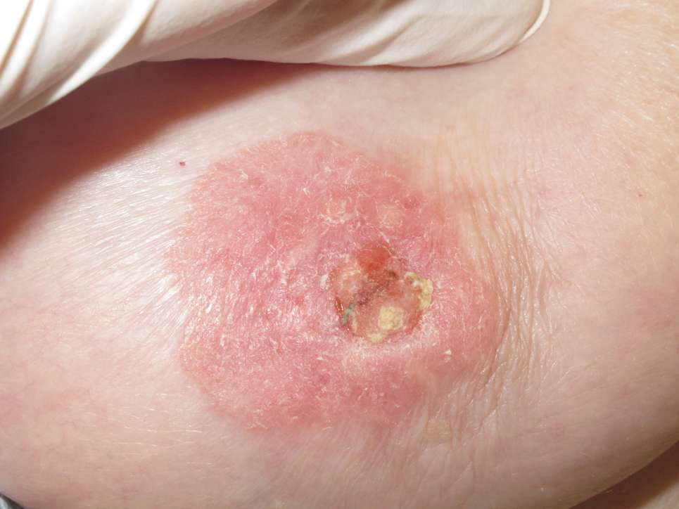
A 91-year old well-appearing white female with history of non-melanoma skin cancer presents with an asymptomatic “rash” on her right nipple of unknown duration. On physical exam, the patient has a broad red patch with adherent scaling involving her right nipple and areola, extending beyond onto the surrounding skin (Figure 1). No palpable mass of the breast nor localized lymphadenopathy were noted on physical exam.
Vote:


ANSWER: A shave biopsy of the right nipple displays distinct intraepidermal cells, with pale cytoplasms and prominent nuclei. Additionally, stains for CAM 5.2 and AE1/AE3 were positive, S100 negative.


Figure 1
Vote:


ANSWER: Paget’s Disease of the Breast


Case Presentation

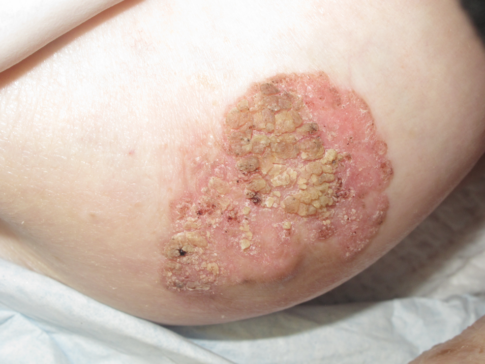
In a second patient, a 50-year-old Caucasian female who presents with this scaly stable stuck-on lesion involving the areola and sparing the nipple of her breast. Parts of it crumble off at times, but then regrow at the base. She has other similar looking lesions on her trunk.
Vote:


ANSWER: Paget’s disease


Case Presentation

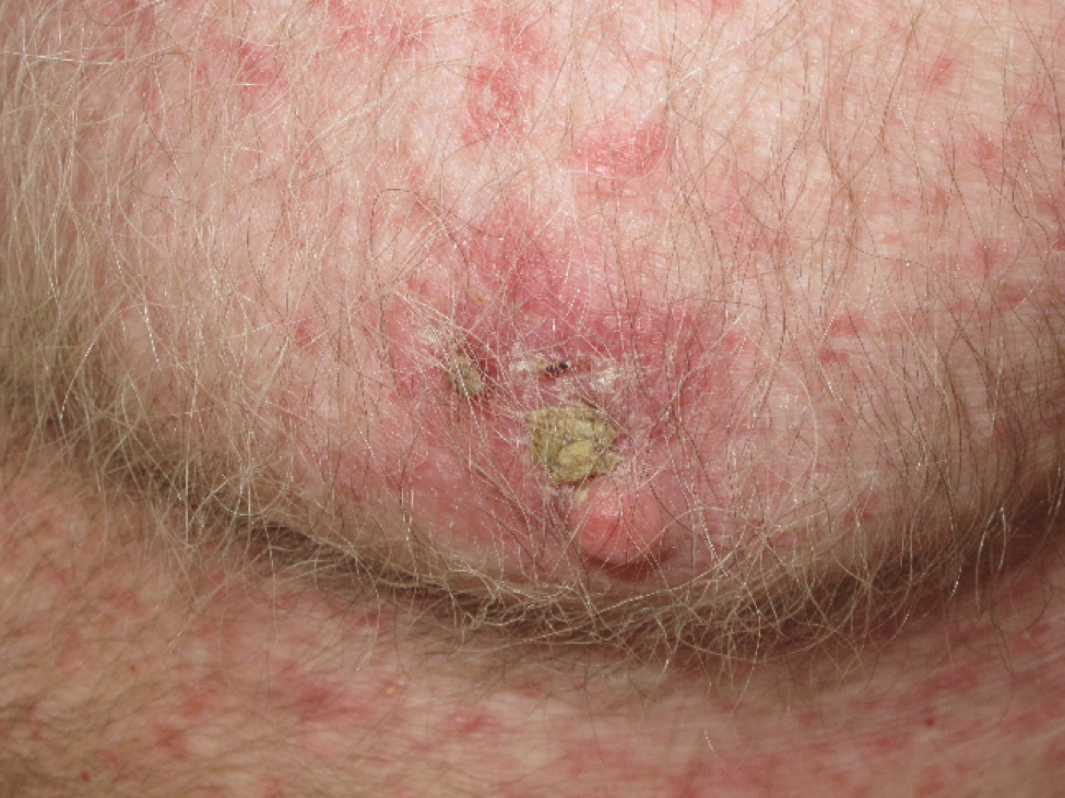
A third patient, a 60-year-old, Caucasian male with history of non-melanoma skin cancer and psoriasis presents with a “rash” involving the skin of the areola eccentrically surrounding but sparing the nipple (figure 3).
A shave biopsy is performed on this patient with histology revealing basaloid cells with relatively uniform hyperchromic cytoplasm in the dermis. The basaloid cells are arranged in lobules with peripheral palisading.
Vote:


ANSWER: Basal cell carcinoma


Discussion
Here, we contrast a classic presentation of an uncommon disease, Paget’s disease of the breast (PDB), and a rare presentation of a common disease, basal cell carcinoma (BCC).
Paget’s disease of the breast is a relatively uncommon skin entity, which often overlies breast cancer. It accounts for a mere 1 to 3% of breast cancers in the United States1. PDB is characterized by a scaly, vesicular, or ulcerated lesion that begins on the nipple and proceeds to spread to the areola2. It displays eczematous symptoms and is frequently associated with symptoms of pain, pruritus, and/or burning. These symptoms can be suggestive of a reactive dermatoses (an eczema) rather than a skin cancer, and therefore warrant special diagnostic care3. Since these lesions can be difficult to distinguish clinically from inflammatory or other neoplastic conditions, a full-thickness shave, wedge or punch biopsy of the nipple is needed in confirming any suspicion of this diagnosis4. One unique feature helpful in distinguishing PDB is its consistent involvement of the nipple, whereas the BCC case presented clinically spared the nipple. The histopathology of PDB is fairly characteristic, showing malignant intraepithelial adenocarcinoma cells with prominent nuclei. Special stains are crucial in distinguishing PDB from melanoma and other tumors. PDB is typically positive for CEA and negative for S-100, whereas melanoma has the opposite findings. Additionally, 84-91% of cases of PDB overexpress HER2.
Paget’s disease of the breast is more common in women and usually affect individuals over age 50. It is generally unilateral, but can present bilaterally5. Greater than 90% of cases are associated with an underlying breast carcinoma, most commonly ductal carcinoma6. Prognosis depends greatly on the size and presence or absence of a palpable breast mass. Five-year survival rates for women with a palpable breast mass range from 20 to 60%, whereas five-year survival is between 75 and 100% for women without palpable breast mass7.
Basal cell carcinoma, in contrast to PDB, is the most common cause of skin cancer, with an estimated incidence of 3 million cases per year in the United States alone. It typically occurs in sun-exposed areas, with approximately 70% occurring on the face, and is only rarely reported involving the nipple8,9. To date there have only been 55 cases of BCC of the nipple and areola complex reported, the majority of cases being in Caucasian men10. Men are theorized to have increased risk due to greater sun exposure of the chest11. Previous history of BCC, immunosuppression, exposure to arsenic, coal tar derivatives and irradiation are also risk factors implicated in BCC; nevertheless, exposure to ultraviolet radiation is the most important risk factor12. Basal cell carcinomas are histologically distinct, exhibiting basaloid cells arranged in lobules in the dermis with relatively uniform hyperchromic cytoplasms and peripheral palisading. BCC is usually diagnostic without the need for special stains, with nodular and superficial BCC subtypes most frequently reported in BCC of the nipple and areola complex13.
Treatment approaches differ for PDB and BCC. Treatment options for PDB depend on the presence of underlying ductal carcinoma and include lumpectomy, excision, radiation, and chemotherapy. Tamoxifen may also be used in patients with an underlying ductal cell carcinoma and estrogen receptor positivity. BCC of the trunk, especially when smaller in size and superficial in nature, can often be managed with desiccation and curettage or excision. However, given the three reported cases of metastatic BCC of the nipple and areola complex, although no formal guidelines are available to guide therapeutic choices, surgical management with Moh’s surgery offers the highest possible cure rate seems preferable10.
It is important to consider comorbidities when managing skin cancer. In a patient with chronic immunosuppression, such as this patient with severe psoriasis on cyclosporine (or a TNF blockers), context is important. If he is growing many skin cancers, or they are growing rapidly, he may be required to come off his immunosuppressive medications, which can increase malignancy growth and aggressiveness. Furthermore, many immunocompromised patients benefit from long term treatment with acitretin (a systemic retinoid FDA approved for psoriasis) which suppresses the growth of many skin cancers.


Post-Test Question:Paget’s/BCC of the nipple


ANSWER: PDB has consistent involvement of the nipple, whereas BCC may spare the nipple
References
1. Berg JW, Hutter RV. Breast cancer. Cancer 1995; 75:257.
2. Franceschini G, Masetti R, D'Ugo D, et al. Synchronous bilateral Paget's disease of the nipple associated with bilateral breast carcinoma. Breast J 2005; 11:355.
3. Chaudary MA, Millis RR, Lane EB, Miller NA. Paget's disease of the nipple: a ten year review including clinical, pathological, and immunohistochemical findings. Breast Cancer Res Treat 1986; 8:139.
4. Gupta RK, Simpson J, Dowle C. The role of cytology in the diagnosis of Paget's disease of the nipple. Pathology 1996; 28:248.
5. Franceschini G, Masetti R, et al.: Synchronous bilateral Paget’s disease of the nipple associated with bilateral breast carcinoma. Breast J.
6. Sakorafas GH, Blanchard K, Sarr MG, Farley DR. Paget's disease of the breast. Cancer Treat Rev.2001;27:9–18.
7. Kollmorgen DR, Varanasi JS, Edge SB, Carson WE 3rd. Paget's disease of the breast: a 33-year experience. J Am Coll Surg 1998; 187:171.
8. American Cancer Society Cancer Facts and Figures 2010. http://www.cancer.org/acs/groups/content/@epidemiologysurveilance/documents/document/acspc-026238.pdf (Accessed on June 10, 2016).
9. Vitasa BC, Taylor HR, Strickland PT, et al. Association of nonmelanoma skin cancer and actinic keratosis with cumulative solar ultraviolet exposure in Maryland watermen. Cancer 1990; 65:2811.
10. Chun KA, Cohen PR. Basal Cell Carcinoma of the Nipple-Areola Complex: A Comprehensive Review of the World Literature. Dermatol Ther (Heidelb) 2016.
11. Shertz WT, Balogh K. Metastasizing basal cell carcinoma of the nipple. Arch Pathol Lab Med. 1986; 110(8):761-2.
12. Huang CW, Pan CK, Tsai CC, et al. Basal cell carcinoma of the nipple-areola complex: a case report. Kaohsiung J Med Sci2005;21:480–2.
13. Crowson AN, Magro CM, Kadin M, et al. Differential expression of bcl-2 oncogene in human basal cell carcinoma. Hum Pathol 1996;27:355–359.
Newsletter
Like what you’re reading? Subscribe to Dermatology Times for weekly updates on therapies, innovations, and real-world practice tips.


