- Acne
- Actinic Keratosis
- Aesthetics
- Alopecia
- Atopic Dermatitis
- Buy-and-Bill
- COVID-19
- Case-Based Roundtable
- Chronic Hand Eczema
- Chronic Spontaneous Urticaria
- Drug Watch
- Eczema
- General Dermatology
- Hidradenitis Suppurativa
- Melasma
- NP and PA
- Pediatric Dermatology
- Pigmentary Disorders
- Practice Management
- Precision Medicine and Biologics
- Prurigo Nodularis
- Psoriasis
- Psoriatic Arthritis
- Rare Disease
- Rosacea
- Skin Cancer
- Vitiligo
- Wound Care
Article
Distribution not random for localized infantile facial hemangiomas
Author(s):
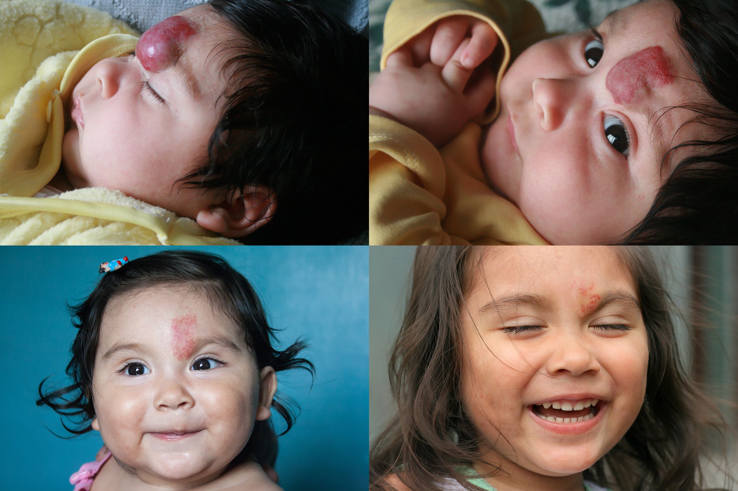
The distribution of localized infantile hemangiomas of the face and scalp favor involvement of the midline face, scalp and ocular axis, shows a retrospective review published in the journal Pediatric Dermatology.
The distribution of localized infantile hemangiomas of the face and scalp favor involvement of the midline face, scalp and ocular axis, shows a retrospective review published in the journal Pediatric Dermatology. (Infantile hemangioma treatment with propranolol. (©AdobeStock,103148250)

The distribution of localized infantile hemangiomas of the face and scalp favor involvement of the midline face, scalp and ocular axis, shows a retrospective review published in the journal Pediatric Dermatology.
“Although previous studies have indicated that localized hemangiomas may have a predilection for sites of fusion between embryologic subunits, including the frontonasal, maxillary, and mandibular prominences, our study did not confirm this. Instead, the hemangiomas occurred most commonly along the ocular axis, which includes the region running horizontally across the eyes and the vertical midline facial axis, with higher densities on the nasal tip and perioral region of the face,” wrote the authors who were led by Anita N. Haggstrom, M.D., of Indiana University. The study was published online August 31.
In an interview with Dermatology Times, Dr. Haggstrom said that many clinicians who see large numbers of patients with infantile hemangiomas, have observed specific commonly affected sites.
“Previous studies had suggested a predilection of localized hemangiomas along the borders of facial developmental segments and it was postulated that perhaps there were local areas of tissue hypoxia at these borders that stimulated hemangioma growth,” Dr. Haggstrom said. “However, our study did not confirm these findings, which was somewhat surprising. The strong predilection for the midline for scalp hemangiomas was likewise unexpected.”
The study concluded that the regions corresponding to the boundaries between the embryonic facial segments, including the maxillary and mandibular metameres, were no more prevalent in the distribution of infantile hemangiomas.
The results also differ from a 2016 study in Pediatric Dermatology that evaluated the distribution of lip hemangiomas, in which upper lip infantile hemangiomas were much more common than lower lip.
“Our study demonstrated a lower lip predilection,” Dr. Haggstrom says. “Conversely, midline hemangiomas of the head and neck are not rare and in fact are a common presentation that should not prompt more extensive workup, which may be reassuring to primary caregivers.”
THE STUDY
Researchers evaluated 4,153 local face and scalp infantile hemangiomas from 10 pediatric dermatology healthcare centers. The hemangiomas were mapped with heat-map software using standardized facial templates displaying frontal and lateral views of the face and frontal, lateral, top and posterior views of the scalp.
71 percent of the hemangiomas were mapped to a frontal facial template, of which 73.8 percent occurred along the midline axis or perpendicularly across the ocular axis in a cross-shaped area of predilection intersecting at the glabella.
Scalp hemangiomas showed a predilection for the midline with 50.5 percent (149 of 295) observed on the top of the scalp at the midline.
Localized hemangiomas did not indicate a preferential laterality.
SIGNIFICANCE
“The study shows novel areas of predilection for infantile hemangiomas, which may eventually help guide our understanding of hemangioma pathogenesis,” Dr. Haggstrom said.
From a developmental prospective, it is unclear why infantile hemangiomas are more frequent at some anatomic sites, despite evidence that infants with these tumors have an increased number of circulating endothelial progenitor cells. “But why these cells would gravitate to particular sites is not known,” she said. “We have also been unable to identify a mechanistic reason based on arterial or other vascular distribution.”
Nonetheless, the results of the current study beg more questions, Dr. Haggstrom said. “Why do these hemangiomas occur in these locations?” she says. “Are there tissue factors specific to these areas that lend just the right environment for hemangioma growth? Ultimately, we would like the study to contribute to our understanding of hemangioma development.”
Dr. Haggstrom and her associates are now looking more closely at other types of infantile hemangiomas and the patterns in which they occur. “We are also trying to identify high-risk versus low-risk patterns to help guide physician management,” she said.
REFERENCE
Haggstrom AN, Baselga E, Chamlin SL, et al. “Localized Infantile Hemangiomas of the Face and Scalp: Predilection for the Midline and Periorbital and Perioral Skin,” Pediatric Dermatology, 2018 Aug 31. DOI:10.1111/pde.13626. [Epub ahead of print]
Yanes DA, Pearson GD, Witman PM. “Infantile Hemaniomas of the Lip: Patterns, Outcomes, and Implications,” Pediatric Dermatology, 2016:33:511-517. DOI:10.1111/pde.12928.
Newsletter
Like what you’re reading? Subscribe to Dermatology Times for weekly updates on therapies, innovations, and real-world practice tips.


