- Acne
- Actinic Keratosis
- Aesthetics
- Alopecia
- Atopic Dermatitis
- Buy-and-Bill
- COVID-19
- Case-Based Roundtable
- Chronic Hand Eczema
- Chronic Spontaneous Urticaria
- Drug Watch
- Eczema
- General Dermatology
- Hidradenitis Suppurativa
- Melasma
- NP and PA
- Pediatric Dermatology
- Pigmentary Disorders
- Practice Management
- Precision Medicine and Biologics
- Prurigo Nodularis
- Psoriasis
- Psoriatic Arthritis
- Rare Disease
- Rosacea
- Skin Cancer
- Vitiligo
- Wound Care
Article
First Case Report of Vitiligo Triggered by Dupilumab Treatment
Author(s):
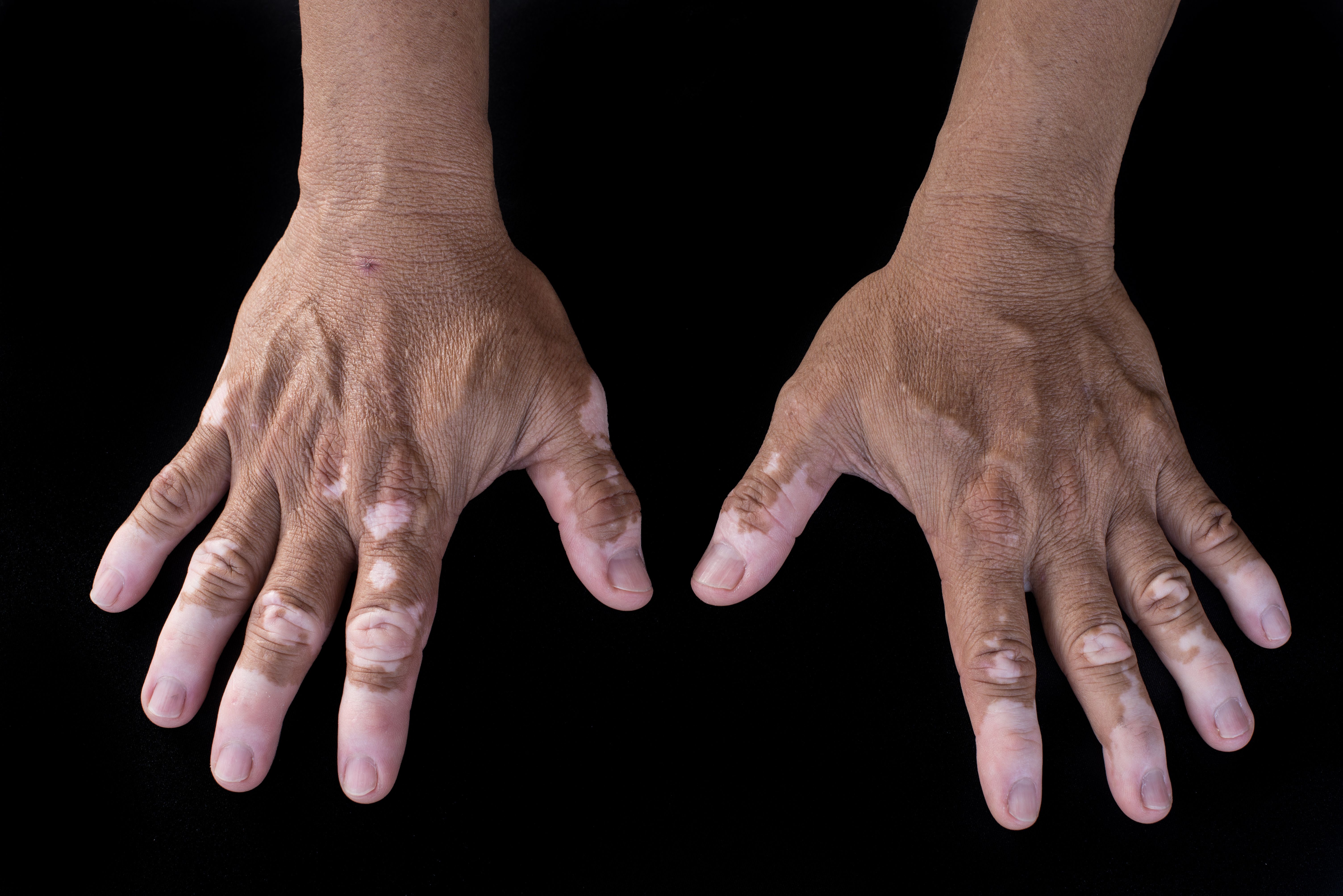
Researchers have identified what may be the first report of vitiligo induced de novo by dupilumab therapy.
In a recent case report,1 researchers identified what may be the first reported case of vitiligo induced de novo by dupilumab therapy in a patient with atopic dermatitis.
anonpichit/AdobeStock

A 79-year-old patient, male, presented to researchers with vitiligo lesions that had developed after dupilumab therapy for his persistent atopic dermatitis. Over the course of his 20 years with the condition, he had been treated with several other therapies, including systemic therapies (including cyclosporine and methotrexate), topical corticosteroids, and narrow band (nb)-UVB phototherapy. These treatments had resulted in temporary or limited signs of disease improvement.
During an initial visit with researchers, the patient had eczematous lesions impacting the back of his hands, as well as his neck and scalp. Researchers established the following baseline scores:
- Dermatology Life Quality Index (DLQI) score of 20
- Eczema Area and Severity Index (EASI) score of 25
- Pruritus-Numeric Rate Scale (P-NRS) score of 9
Due to these scores, researchers began treating the patient with an initial dose of 600 mg of dupilumab, followed by 300 mg of the drug delivered every 2 weeks. After 4 weeks of treatment, researchers determined the patient had entered complete atopic dermatitis remission, establishing the following scoring:
- DLQI score of 0
- EASI score of 0
- P-NRS score of 0
However, researchers noted that the patient had hypopigmented macules impacting the regions where eczematous lesions had once been. They conducted a Wood’s lamp examination and a skin biopsy.
They found several achromic patches, as well as an absence of melanocytes in the epidermis’ basal layer and dermal inflammatory infiltrate of lymphocytes, with the latter having notable perivascular distribution.
Upon diagnosing the patient with vitiligo, researchers continued to treat the patient with dupilumab, in addition to beginning treatment with topical corticosteroids and nb-UVB phototherapy. 16 weeks later, they found the patient had undergone complete remission of vitiligo and atopic dermatitis, again with the following established scoring:
- DLQI score of 0
- EASI score of 0
- P-NRS score of 0
While researchers note this is currently the first report of such a case in the literature of vitiligo, they cited 1 existing publication2 wherein researchers described a patient with atopic dermatitis presenting with an enlarged achromic patch of vitiligo after beginning treatment with dupilumab.
“Our case appears to be currently the first report in the literature of vitiligo induced de novo by dupilumab therapy. The molecular drivers of vitiligo occurring during dupilumab treatment are unclear. The pathogenetic mechanism could be related to the imbalance between the T helper(Th)2 and Th1/Th17 pathways,” case report authors wrote. “However, new studies on this topic are needed to better understand the pathophysiological mechanisms underlying this rare AE [adverse event] during dupilumab therapy.”
References
- Picone V, Napolitano M, Torta G, Fabbrocini G, Patruno C, Vitiligo during dupilumab therapy: a case report., JAAD Case Reports (2023), doi: https://doi.org/10.1016/ j.jdcr.2023.03.025.
- Takeoka S, Kamata M, Yokoi I, Takehara A, Tada Y. Rapid enlargement of vitiligo vulgaris after journal pre-proof initiation of dupilumab for atopic dermatitis: a case report. Acta Derm Venereol. 2021 Oct 110 28;101(10):adv00581.
Newsletter
Like what you’re reading? Subscribe to Dermatology Times for weekly updates on therapies, innovations, and real-world practice tips.


