- Acne
- Actinic Keratosis
- Aesthetics
- Alopecia
- Atopic Dermatitis
- Buy-and-Bill
- COVID-19
- Case-Based Roundtable
- Chronic Hand Eczema
- Chronic Spontaneous Urticaria
- Drug Watch
- Eczema
- General Dermatology
- Hidradenitis Suppurativa
- Melasma
- NP and PA
- Pediatric Dermatology
- Pigmentary Disorders
- Practice Management
- Precision Medicine and Biologics
- Prurigo Nodularis
- Psoriasis
- Psoriatic Arthritis
- Rare Disease
- Rosacea
- Skin Cancer
- Vitiligo
- Wound Care
News
Article
Injected Hyaluronic Acid Stimulates Collagen Production in Photoaged Skin
Author(s):
An investigation into skin changes at the cellular level provides insight into hyaluronic acid’s benefits.
blackday/AdobeStock

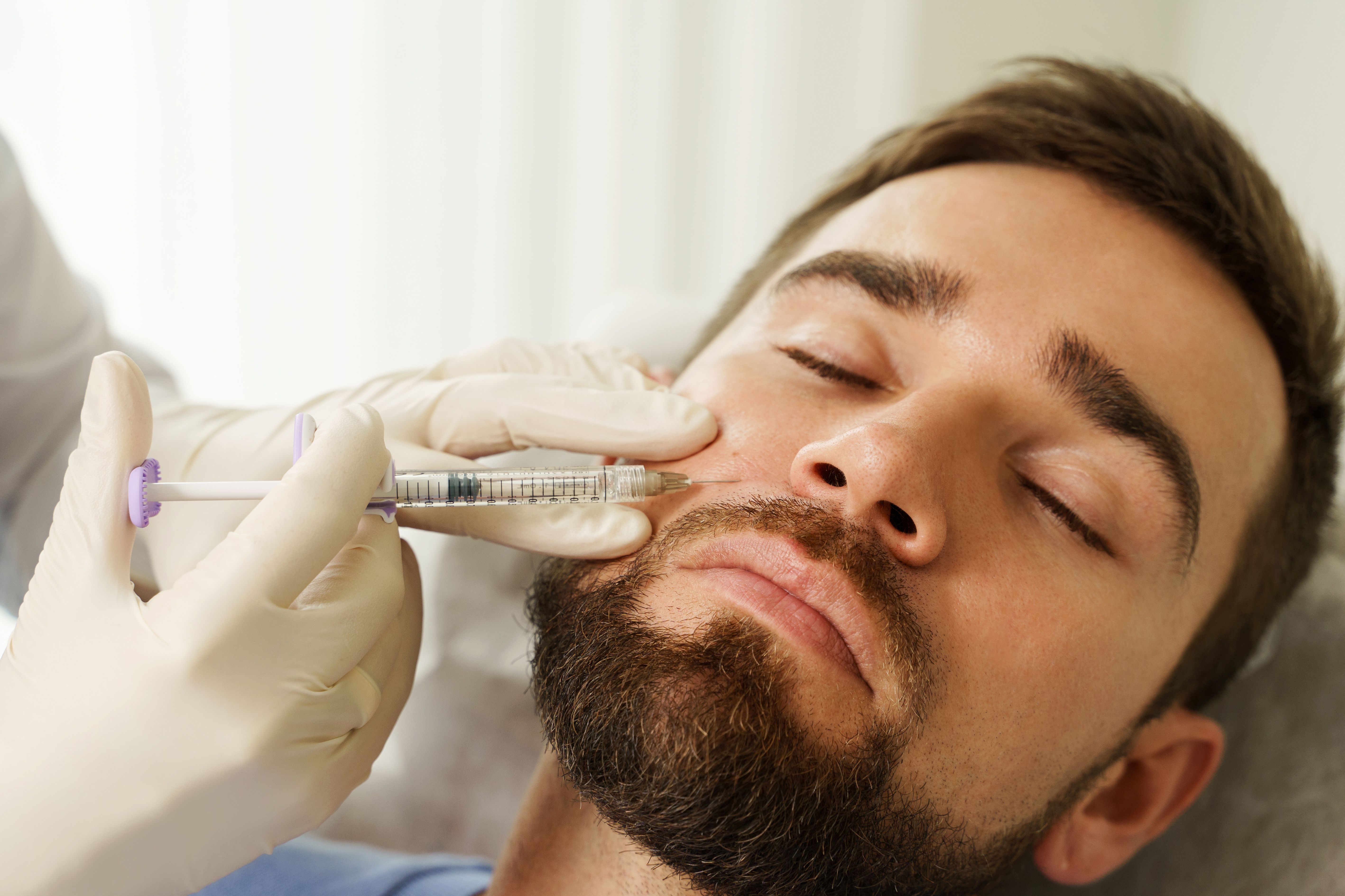
Cross-linked hyaluronic acid dermal filler not only adds volume to the skin, but also aids in collagen production, researchers at the University of Michigan Medical School have found.1
Professor of clinical dermatology Frank Wang, MD, et al study the mechanisms behind skin photoaging and the effects of cross-linked hyaluronic acid (CL-HA) fillers.
They have observed in prior studies that injection of CL-HA dermal fillers into photoaged skin could reverse changes to type 1 collagen bundles and fibroblasts by stimulating the fibroblasts to adopt a stretched morphology and synthesize type I procollagen.2
This fibroblast stimulation results from enhanced structural support and mechanical forces induced by the filler in the surrounding dermal extracellular matrix, Wang et al found.
For the group’s newest study, they injected CL-HA and vehicle (saline) into photodamaged forearm skin of subjects older than 60 years and took skin samples at various timepoints.
Punch biopsies of 4 mm were obtained under local anesthesia (lidocaine) at 1, 2, 3 and 4 weeks post-injection for an early group of subjects (15/16 completed the study), and 3, 6, 9 and 12 months for a late group (16/18 completed the study).
Evaluating the biopsies, the group “investigated when fibroblast elongation and activation begin following CL-HA injection, and the duration and mechanisms of these changes,” they reported, and evaluated whether fibroblast activation leads to deposition of type I collagen.
Prior studies had not specifically investigated whether newly synthesized type I procollagen is converted to intact type I collagen bundles, they wrote.
Study Results
In vehicle-injected skin, Wang et al reported, collagen bundles “appeared shortened/fragmented, thin, loosely packed and separated from each other.”
“As early as 4 weeks in filler-injected skin, collagen bundles appeared thick and densely packed, particularly around dermal pockets containing injected filler. Compared with vehicle-injected skin, these bundles appeared bright, suggesting that they were comprised of compactly arranged type I collagen fibrils.”
Additionally, in filler-injected skin at 12 months, second-harmonic generation imaging revealed numerous bright collagen bundles, which appeared long, thick, and densely packed in the extracellular matrix (ECM), they reported.
Clinically, these results mean that immediately following CL-HA injection, skin volumization is likely “derived from physical presence of the filler, which enhances mechanical support (and fibroblast elongation) by occupying space in the dermal ECM. Within 1 month, deposition of long, thick, densely packed, intact collagen bundles likely provides additional mechanical support, enhancing early clinical improvement and fibroblast elongation,” they wrote.
“At 12 months,” they added, “substantial amounts of the injected filler remained present, suggesting that the clinical benefits of injected CL-HA are long-lasting due to both collagen deposition and space occupation by the filler.”
Therefore, single injections of CL-HA fillers “can lead to rapid and long-lasting improvement of skin by stimulating collagen deposition,” and “repeat injections may add more collagen, eventually reducing the need for re-treatment," said Wang in a University of Michigan press release.3
References
- Wang F, Do TT, Smith N, et al. Implications for cumulative and prolonged clinical improvement induced by cross-linked hyaluronic acid: An in vivo biochemical/microscopic study in humans. Exp Dermatol. 2024;33(1):e14998. doi:10.1111/exd.14998
- Quan T, Wang F, Shao Y, et al. Enhancing structural support of the dermal microenvironment activates fibroblasts, endothelial cells, and keratinocytes in aged human skin in vivo. J Invest Dermatol. 2013; 133(3): 658-667. doi:10.1038/jid.2012.364
- Roy T. Michigan Medicine. Turning back the clock on photoaging skin. News release. February 14, 2024. Accessed February 15, 2024. https://www.michiganmedicine.org/health-lab/turning-back-clock-photoaging-skin
Newsletter
Like what you’re reading? Subscribe to Dermatology Times for weekly updates on therapies, innovations, and real-world practice tips.


