- Acne
- Actinic Keratosis
- Aesthetics
- Alopecia
- Atopic Dermatitis
- Buy-and-Bill
- COVID-19
- Case-Based Roundtable
- Chronic Hand Eczema
- Chronic Spontaneous Urticaria
- Drug Watch
- Eczema
- General Dermatology
- Hidradenitis Suppurativa
- Melasma
- NP and PA
- Pediatric Dermatology
- Pigmentary Disorders
- Practice Management
- Precision Medicine and Biologics
- Prurigo Nodularis
- Psoriasis
- Psoriatic Arthritis
- Rare Disease
- Rosacea
- Skin Cancer
- Vitiligo
- Wound Care
Article
Laser technology creates hi-res maps of acne lesions
Author(s):
An exploration of the feasibility of using laser technology to detect changes in microvasculature during the acne lesion healing progress and to introduce a microvasculature-based biomarker in evaluating the treatment and grading of acne.
The findings of a new study may help foster a better understanding of acne and its subtypes-which in turn could lead to more effective ways to compare and improve the therapeutic efficacy of treatment products.
Changes in microvasculature
Researchers from the University of Washington and Yildirim Beyazit University in Ankara, Turkey, designed a study to explore the feasibility of using a type of laser technology to detect changes in microvasculature during the acne lesion healing progress and to introduce a microvasculature-based biomarker in evaluating the treatment and grading of acne.
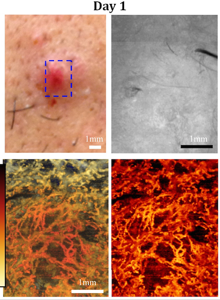
According to study author Ruikang K Wang, Ph.D., professor of bioengineering and ophthalmology at the University of Washington, he and his colleagues developed a non-invasive imaging technology that can three-dimensionally map functional vascular-network innervating living tissue, at the level of capillaries, by “merely shining a light” onto the tissue.
NEXT: Optical coherence tomography and day 3 photos
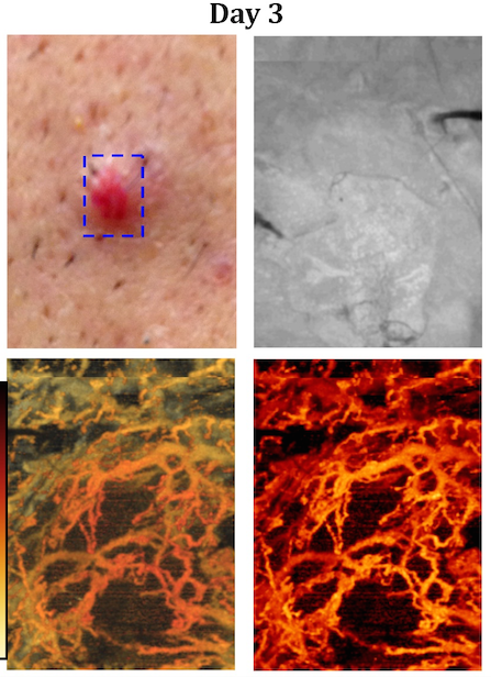
“The technique is based on optical coherence tomography (OCT), a popular technology clinically adopted in ophthalmology,” Dr. Wang tells Dermatology Times. “It is extremely sensitive to moving particles, thus providing us an opportunity to contrast moving blood cells within vessels without the use of any contrast agents. We call this technology optical microangiography (OMAG).”
The system operates on a central wavelength of 1,310 nanometers with an A-line rate of 100 kilohertz and has an extended imaging range. Using handheld probes, researchers scanned acne lesions on volunteer patients’ facial skin to obtain images. They tested the technology for longitudinal imaging of changes in microvasculature and structure during acne lesion development and developed a microvasculature-based biomarker to aid in the treatment and the assessment of the acne’s severity.
NEXT: Clinicians employ various methods
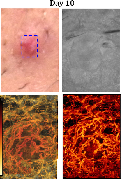
“Currently, clinicians employ various methods to assess the severity of acne,” Dr. Wang says. “However, no single grading system has been universally accepted. We believe new research tools are needed to better understand the natural history, subtypes and triggers of acne, and to improve and compare treatment options. The ability of OMAG to delineate functional microvascular network, at the level of capillary vessels, can be very useful in facilitating the clinical treatment of acne lesion scarring.
“We know that laser-based acne-treatment methods modify the subdermal microvasculature,” he adds. “In this case, OMAG can play an important role in guiding and improving these treatment methods.” Vascular density as a biomarker.
NEXT: Vascular density in acne
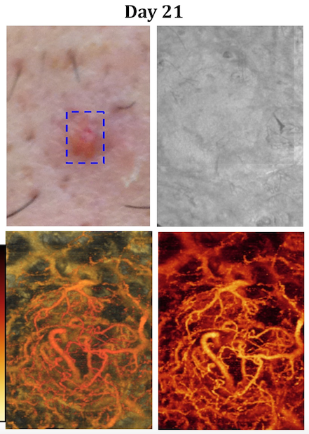
According to the study, vascular density in the acne lesion can be used as a biomarker for grading acne severity and the assessment of the effectiveness of the treatments.
“For example, instead of applying irritating and potentially dangerous topical drugs on a larger portion of a patient’s skin, drugs can be applied to a small area to see the drug’s effectiveness on a specific patient,” Dr. Wang says.
NEXT: Density derived from non-invasive OMAG
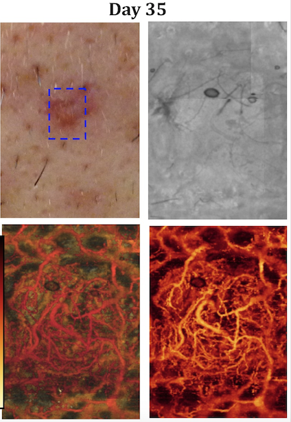
He adds that the vascular density derived from non-invasive OMAG measurement may also be used as a biomarker in the monitoring, therapeutic treatment and management of other skin diseases, such as port wine stain (PWS), psoriasis, rosacea, skin burn and skin cancer.
NEXT: Vascular malfunction in superficial dermal capillaries in PWS
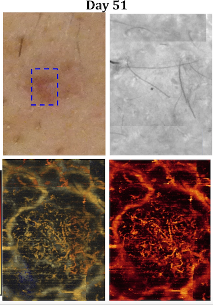
“For instance, vascular malfunction in superficial dermal capillaries in PWS can easily be characterized with vascular density derived from OMAG,” Dr. Wang says. “Moreover, as a replacement for a standard visual inspection of the skin burn, the scar’s vascular density can be a potential indicator to assess scar progression, providing diagnosis and proper treatment of pathological scarring.”
The study was published in the journal Lasers in Surgery in Medicine.
Disclosures:
Dr. Wang has a financial interest in patents filed by the Oregon Health & Science University (OHSU) and the University of Washington (UW) for the OMAG technology. This potential individual conflict of interest has been reviewed and managed by OHSU and UW.
Newsletter
Like what you’re reading? Subscribe to Dermatology Times for weekly updates on therapies, innovations, and real-world practice tips.


