- Acne
- Actinic Keratosis
- Aesthetics
- Alopecia
- Atopic Dermatitis
- Buy-and-Bill
- COVID-19
- Case-Based Roundtable
- Chronic Hand Eczema
- Chronic Spontaneous Urticaria
- Drug Watch
- Eczema
- General Dermatology
- Hidradenitis Suppurativa
- Melasma
- NP and PA
- Pediatric Dermatology
- Pigmentary Disorders
- Practice Management
- Precision Medicine and Biologics
- Prurigo Nodularis
- Psoriasis
- Psoriatic Arthritis
- Rare Disease
- Rosacea
- Skin Cancer
- Vitiligo
- Wound Care
News
Article
LC-OCT Demonstrates Potential For Rapid, Effective Visualization of Inflammatory Conditions Across Skin Types
Author(s):
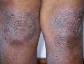
LC-OCT provided quick, noninvasive assessment of skin conditions with detailed imaging aids diagnosis and treatment, especially for patients with darker skin types.
Line-field confocal optical coherence tomography (LC-OCT) demonstrated rapid and effective visualization of inflammatory skin conditions, particularly atopic dermatitis, in patients of varying skin types, according to a study published in the Journal of the European Academy of Dermatology and Venereology.1
Noninvasive imaging technologies such as reflectance confocal microscopy (RCM) and LC-OCT offer a detailed look at the microscopic features of atopic dermatitis without the need for biopsies and the scarring they can cause. RCM technology, for example, has revealed characteristic features of atopic dermatitis, such as acanthosis and spongiosis, visible on histopathology.2
LC-OCT combines the cellular resolution of RCM with the imaging depth of optical coherence tomography, producing 3D images. While RCM provides high-resolution images parallel to the skin surface, LC-OCT offers the capability to capture images in both horizontal and vertical planes simultaneously. This, study authors noted, led them to investigate its efficacy and speed in the visualization of inflammatory skin conditions, especially among patients with atopic dermatitis.
The prospective cohort study, conducted at Rutgers Center for Dermatology, involved 10 patients with clinically diagnosed atopic dermatitis. Patients were examined on both lesional and perilesional skin using LC-OCT and RCM.
Image acquisition was performed using LC-OCT and RCM, capturing both horizontal and vertical images of lesional and perilesional skin. Researchers conducted quantitative analysis to measure epidermal thickness and dermo-epidermal junction undulation, while visually analyzing microscopic features such as spongiosis, exocytosis, and perivascular inflammation.
Their analysis of epidermal thickness revealed significant differences between lesional and healthy skin, with atopic dermatitis lesions exhibiting thicker stratum corneum and increased living and total epidermal thickness.
Furthermore, LC-OCT identified characteristic features of atopic dermatitis, including spongiosis, exocytosis, and perivascular inflammation, comparable to findings on RCM. Additionally, some features were observed in clinically normal skin, suggesting subclinical alterations in unaffected areas.
To the knowledge of researchers, this study represents the first to examine the use and efficacy of LC-OCT in assessing microscopic pathological features of AD.
"This study supports the potential of LC-OCT for assessing inflammatory skin pathologies. The ability of this technology to be used to visualize microscopic features of AD lesions, including acanthosis, exocytosis, spongiosis, inflammatory infiltrate, and atypical blood vessels, lends to its potential utility as a fast, noninvasive tool for both diagnosis and treatment monitoring," wrote study authors Ouellette et al. "This may be especially important in patients with darker skin types, as it may elucidate the extent of inflammation in the skin that may not be easily gauged using current clinical severity tools that use redness as a component, such as the EASI. Additionally, LC-OCT may be used to highlight underlying inflammation occurring in normal-appearing skin of patients with atopic dermatitis."
References
- Ouellette S, Truong TM, Khan S, Raji S, Razi S, Rao B. Assessment of atopic dermatitis in all phototypes using line-field confocal optical coherence tomography (LC-OCT) and reflectance confocal microscopy (RCM). J Eur Acad Dermatol Venereol. April 16, 2024. Accessed April 19, 2024. https://doi.org/10.1002/jvc2.421
- Bratu D, Boda D, Caruntu C. Reflectance confocal microscopy in monitoring atopic dermatitis treated with topical calcineurin inhibitors. Healthcare. 2023. Accessed April 22, 2024. https://doi.org/10.3390/healthcare11020152
Newsletter
Like what you’re reading? Subscribe to Dermatology Times for weekly updates on therapies, innovations, and real-world practice tips.


