- Acne
- Actinic Keratosis
- Aesthetics
- Alopecia
- Atopic Dermatitis
- Buy-and-Bill
- COVID-19
- Case-Based Roundtable
- Chronic Hand Eczema
- Chronic Spontaneous Urticaria
- Drug Watch
- Eczema
- General Dermatology
- Hidradenitis Suppurativa
- Melasma
- NP and PA
- Pediatric Dermatology
- Pigmentary Disorders
- Practice Management
- Precision Medicine and Biologics
- Prurigo Nodularis
- Psoriasis
- Psoriatic Arthritis
- Rare Disease
- Rosacea
- Skin Cancer
- Vitiligo
- Wound Care
News
Article
New Imaging System Differentiates Between Melanoma and Nonmelanoma Nevi with 100% Sensitivity
Author(s):
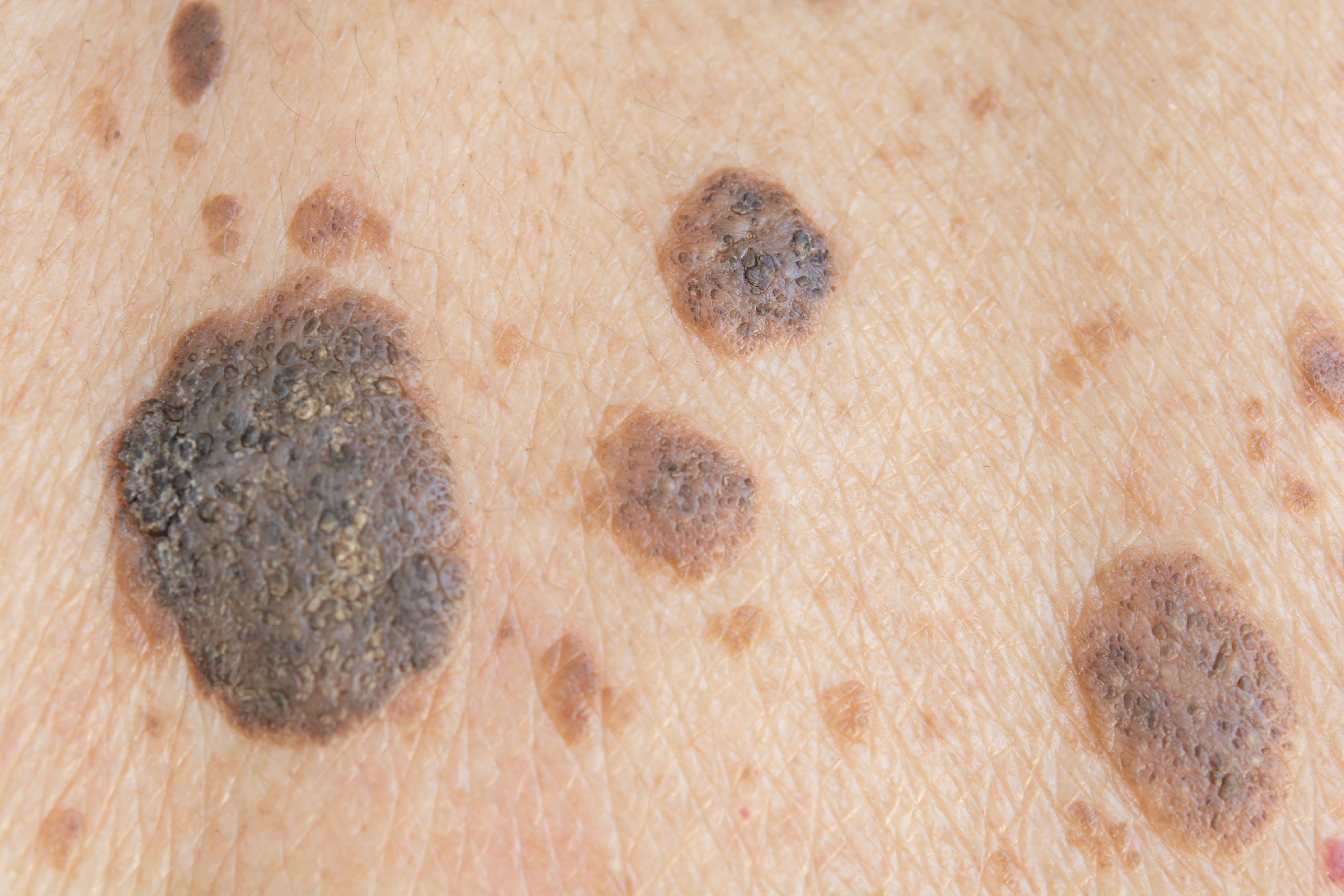
A hand-held skin fluorescence imaging system shows promise in its ability to differentiate between concerning and non-concerning moles.
Srisakorn/AdobeStock

Researchers have developed a new imaging technique—in vivo molecular skin fluorescence imaging (mSFI)—and reported in a recent journal communication that it is able to differentiate melanomas from nonmelanoma nevi with 100% sensitivity and 95.7% specificity.1
The mSFI system can identify dysplastic nevi with the potential for melanoma development, investigators claim, by assessing the presence of tissue remodeling activity.
The results of a study investigating the imaging system, performed by researchers at Orlucent, Inc., appeared in the January 2024 issue of the journal Pigment Cell and Melanoma Research.
“By imaging cells and their crosstalk with the environment, the mSFI provides physicians with the ability to identify lesions at earlier stages than existing technologies,” the authors wrote.
The mSFI system uses a topically applied fluorescent peptide (ORL-1) dye that binds to αvβ3 integrin. It is performed noninvasively using a hand-held device.
Using mSFI, the researchers examined 78 nevi in a pilot clinical study. According to the report, each nevus and its surrounding skin were prepared with a preconditioning solution followed by application of the dye. After a 4-minute incubation period, excess non-bound dye was removed, then photographic and fluorescent images of the mole were captured.
These images were then transferred to a computer for further analysis. The nevi were excised and submitted for biopsy. Histopathological diagnoses were used as the gold standard for the development and evaluation of the image analysis algorithm, the researchers wrote.
Of the 78 nevi, 44 were confirmed by biopsy and histopathology as benign, 26 as atypical or dysplastic, and 8 as melanoma. mSFI differentiated melanomas from non-melanomas with a sensitivity of 100% (8 of the 8 melanomas tested positive) and specificity of 95.7% (67 of the 70 non-melanoma nevi tested negative), according to the report.2
The researchers expressed optimism about the future of mSFi technology, stating it has the potential to “minimize reliance on visual, subjective or invasive tools such as biopsy while allowing the physician to examine biological activity not visible with the naked eye.”
Catherine Shachaf, PhD, the corresponding author of the study and president of Orlucent, stated in a press release, "Incorporating in vivo molecular imaging into clinical management opens new doors to objectively identifying suspicious moles with the potential of becoming melanoma or melanomas with the potential to be aggressive at the earliest stage.
“Simultaneously, it can also help alleviate the anxiety linked to the uncertainty of conventional highly subjective diagnostic methods."
References
- Orlucent. Orlucent demonstrates accurate, non-invasive detection in vivo of melanoma-related activity using first-in-kind handheld mole imaging system. PR Newswire. Published January 16, 2024. Accessed January 16, 2024.https://www.prnewswire.com/news-releases/orlucent-demonstrates-accurate-non-invasive-detection-in-vivo-of-melanoma-related-activity-using-first-in-kind-handheld-mole-imaging-system-302035317.html
- Shachaf A, Manbeck K, Yang G, Shachaf C. (2024). Molecular skin fluorescence imaging: A tool for evaluating early melanoma development. Pigment Cell & Melanoma Research, 00, 1–6. https://doi.org/10.1111/pcmr.13159
Newsletter
Like what you’re reading? Subscribe to Dermatology Times for weekly updates on therapies, innovations, and real-world practice tips.


