- Acne
- Actinic Keratosis
- Aesthetics
- Alopecia
- Atopic Dermatitis
- Buy-and-Bill
- COVID-19
- Case-Based Roundtable
- Chronic Hand Eczema
- Chronic Spontaneous Urticaria
- Drug Watch
- Eczema
- General Dermatology
- Hidradenitis Suppurativa
- Melasma
- NP and PA
- Pediatric Dermatology
- Pigmentary Disorders
- Practice Management
- Precision Medicine and Biologics
- Prurigo Nodularis
- Psoriasis
- Psoriatic Arthritis
- Rare Disease
- Rosacea
- Skin Cancer
- Vitiligo
- Wound Care
Article
New In Vitro Technique Capable of Detecting Skin Wrinkles
Author(s):
Researchers indicated no existing strategy for detecting skin wrinkles in vitro.
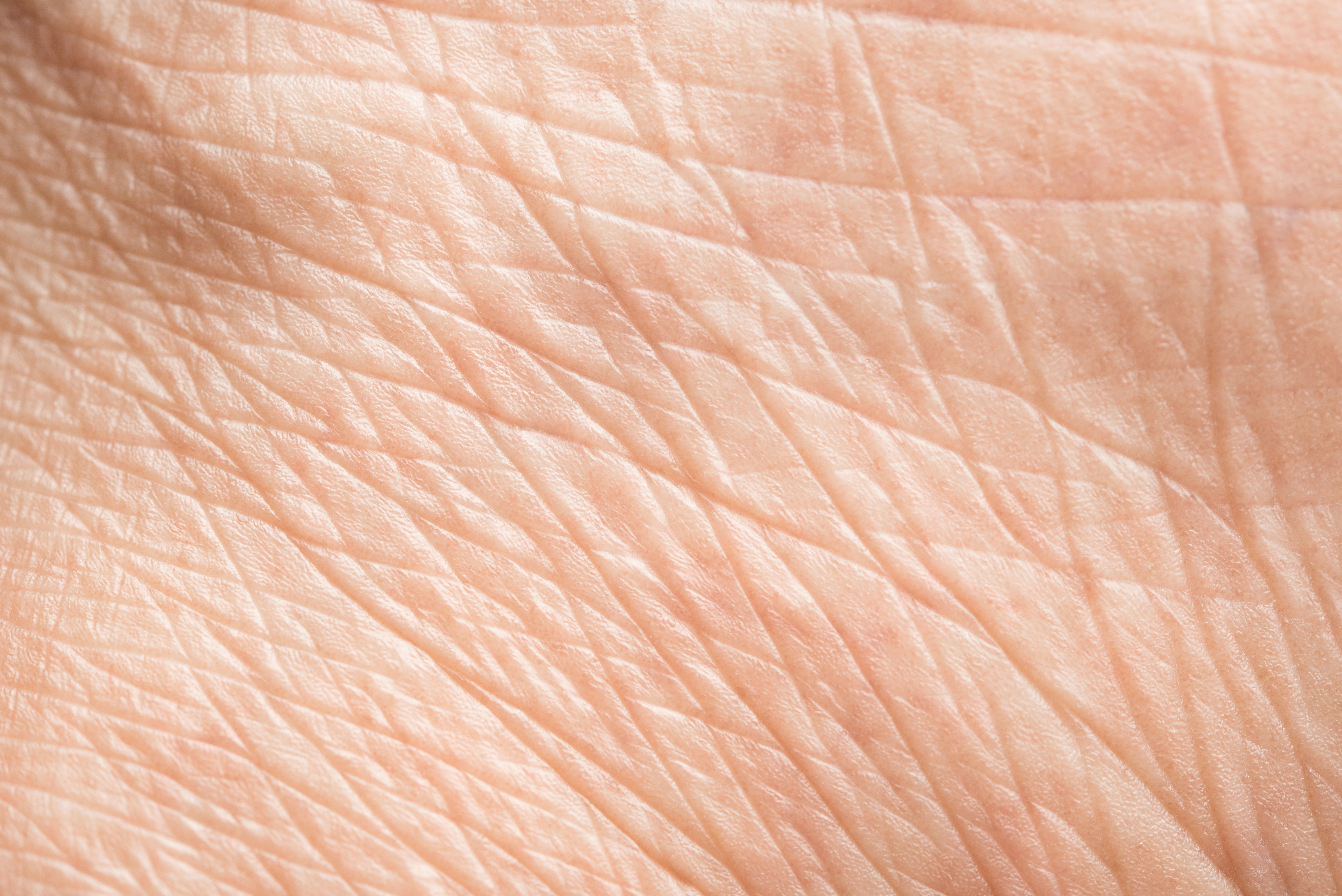
Researchers have developed a novel in vitro technique for identifying skin wrinkles.
Love the wind/AdobeStock

In a study,1 researchers sought to develop a technique, citing a lack of in vitro techniques capable of detecting skin wrinkles, further noting that clinical tests are the only existing method to detect anti-wrinkle efficacy.
They began by conducting a cell culture of human neonatal dermal fibroblasts (HDFn) and human neonatal epidermal keratinocytes (HEKn). Cells were cultured with an Epilife medium, low-serum growth supplements, 100 U/mL of penicillin, and 100 mg/mL of streptomycin at a consistent temperature and in a consistent atmosphere.
Using a 3D-printed holder and mold, researchers were able to mimic the creation of wrinkles in the dermal layer of the skin, particularly during the collagen gelation process, in a new method called collagen stamping.
They then reconstructed full-thickness skin wrinkle mimics by seeding HEKns on the prepared dermis and conducting an air-liquidinterface culture, thus constructing an epidermis. Ultimately, 2 types of skin wrinkle mimics were produced, each with different depths and the same width—one of these was a non-stamped skin mimic.
Retinoic acid treatment was used to examine changes in wrinkles among the skin mimics.
In vivo Optical Coherence Tomography (OCT) imaging and image processing were used to qualify skin wrinkles on the skin equivalents. Using more OCT imaging, researchers also conducted swept-source OCT (SS-OCT) imaging of eye wrinkles on patient volunteers.
They found that, “Unlike natural human skin, the upper dermis of the reconstructed skin model had a flat shape without rete-ridge structures. However, the dermis processed through collagen stamping specifically had a furrow formed consistently with the substructure of the mold."
Furthermore, through SS-OCT imaging, researchers measured the depth of dermal furrows and found that depth had a distinct, positive correlation with the dimensions of the molds.
“The 2D SS-OCT image analyses revealed that the depth and width of wrinkles increased significantly proportionately to the variable length of the mold,” study authors wrote. “In contrast, a significant decrease in the depth and width of the wrinkles was observed in the RA-treated group. These results were confirmed by quantifying the overall wrinkle area.”
Additionally, researchers found that retinoic acid treatment decreased the depth and width of wrinkles that had formed, at a statistically significant rate.
According to researchers, a potential study limitation was the study’s mimicking of thick skin wrinkles as opposed to thin skin wrinkles.
“With the advancement of collagen stamping technology, it will be necessary to diversify the wrinkle models,” study authors wrote. “Furthermore, the clinical correlation should also be verified through clinical evaluation. With these improvements, we expect to develop a more effective substance to alleviate skin wrinkles by increasing the clinical correlation of in vitro skin anti-aging research.”
Reference
- Min D, Ahn Y, Lee HK, Jung W, Kim H. A novel optical coherence tomography‐based in vitro method of anti‐aging skin analysis using 3D skin wrinkle mimics. Skin Res Technol. 2023;29(6). doi:10.1111/srt.13354
Newsletter
Like what you’re reading? Subscribe to Dermatology Times for weekly updates on therapies, innovations, and real-world practice tips.


