- Acne
- Actinic Keratosis
- Aesthetics
- Alopecia
- Atopic Dermatitis
- Buy-and-Bill
- COVID-19
- Case-Based Roundtable
- Chronic Hand Eczema
- Drug Watch
- Eczema
- General Dermatology
- Hidradenitis Suppurativa
- Melasma
- NP and PA
- Pediatric Dermatology
- Pigmentary Disorders
- Practice Management
- Precision Medicine and Biologics
- Prurigo Nodularis
- Psoriasis
- Psoriatic Arthritis
- Rare Disease
- Rosacea
- Skin Cancer
- Vitiligo
- Wound Care
Article
Skin clues help identify cancer metastases
Because skin malignancies often herald internal or systemic malignancies, knowing the link between primary and metastatic sites can impact staging, prognosis and management – and in some cases, save lives.

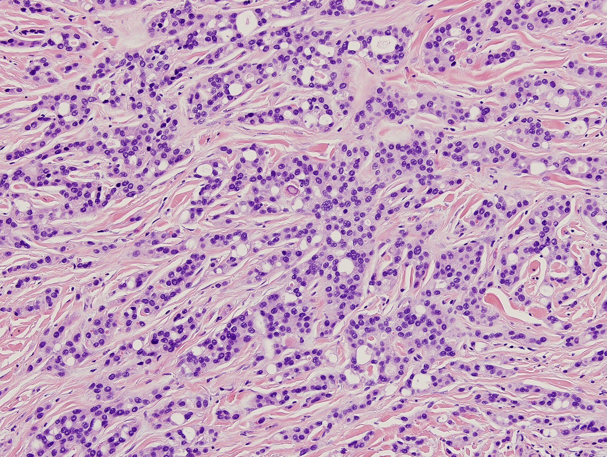
Histopathology of cutaneous metastatic breast carcinoma revealing ductular tumor islands dissecting through dermis. (Photos courtesy of Dr. Vincent Liu)The complex interplay between the skin and systemic malignancies can require dermatologists to use everything from clinical clues to special immunohistochemical stains in identifying primary tumors, according to Vincent Liu, M.D., clinical associate professor, departments of dermatology and pathology, University of Iowa Hospitals & Clinics.
READ: Opportunities, challenges with new treatments for advanced cancer
"The intersection between the skin and systemic malignancy attracted many of us dermatologists to the field – using the skin as a reflection of what's underneath. As I tell our medical students, skin is a window to the soul," Dr. Liu says.
Melanoma is one of the most common metastases encountered in dermatology, he says.
"When it does metastasize, skin is one of the most common targets." In fact, he says that when malignant melanoma metastasizes, it is estimated that half the time it will end up in the skin or subcutaneous tissue. It's important to recognize and diagnose cutaneous metastatic melanoma because this information impacts the patient's staging, prognosis and management, he adds.
One case shared at the 73rd Annual Meeting of the American Academy of Dermatology (San Francisco, 2015) involved breast cancer that had metastasized to the skin, resulting in an indurated, erythematous plaque on the patient's upper arm.
"The biopsy from that lesion disclosed typical carcinoma cells infiltrating the dermis. The relatively high incidence of breast carcinoma in the U.S. population compels dermatologists to consider it in their differential diagnosis," Dr. Liu says. Breast carcinoma metastases generally favor the anterior chest wall and abdomen.
NEXT: Unknown primary tumors
Unknown primary tumors

Discrete ovoid plaque on shoulder representing metastasis to skin of a primary breast carcinoma. (Photos courtesy of Dr. Vincent Liu)"In some cases, a patient will present with a metastatic skin lesion without a known primary origin," which occurs in an estimated 4.4% of all cutaneous metastases.1
In such cases, "Identifying the primary tumor can be the key question" to guide management; however, diagnosing such tumors pose many challenges.
READ: Dermatologists role in diagnosing, managing advanced BCC
First, says Dr. Liu, several other tumors and even inflammatory lesions can mimic cutaneous metastases both clinically and microscopically. Examples include keratinocytic, vascular and melanocytic processes.
Moreover, "Distinction of a skin metastasis from a primary skin tumor often proves difficult. Prognosis in general depends very much on whether the tumor is a primary or secondary tumor. Clinically, and particularly pathologically, however, we'll occasionally encounter tumors where one can't be entirely certain if it represents a primary skin tumor, or if it's a metastasis from somewhere else," he says.
In those cases believed to represent metastatic disease, he says, establishment of the primary origin can still be problematic, especially given the de-differentiation that such tumors can undergo. In these instances, the analysis can be partly informed by knowledge of relative frequencies of skin metastasis by different tumors, which is a function of how commonly the primary tumor occurs overall, how likely it is to metastasize, and how commonly its metastases target the skin, Dr. Liu says. Additionally, "Different primary tumors have certain predilections for targeting specific anatomic sites." For example, he says, if a skin metastasis appears on the chest, statistics point toward a primary breast or lung tumor.
Immunohistochemistry using special stains such as S100 (which stains positive in melanoma) also helps in challenging cases, Dr. Liu says.
"But even immunohistochemical panels are not 100% certain. That's where integration of the entire clinicopathologic picture is crucial," he says.
NEXT: Systemic malignancies
Systemic malignancies

Seemal R. Desai, M.D.Hematolymphoid malignancies offer unique interactions with skin. Dr. Liu says these malignancies are important not only because they can involve the skin directly, but they're also commonly associated with paraneoplastic dermatoses.
READ: Fungal infections may mimic malignancies
Seemal R. Desai, M.D., adds, "Skin conditions are often the heralding sign of a more serious cancer inside the body. We must be in tune with our patients because we potentially save lives by linking a skin condition to an underlying cancer." He is founder and medical director of Innovative Dermatology, P.A., in Plano, Texas, and clinical assistant professor of dermatology at the University of Texas Southwestern Medical Center.
One case Dr. Desai presented involved a 70-year-old male with a six-month history of thickened, enlarging plaques that started on the hands and progressed to the lower body. Biopsy showed psoriasiform epidermal hyperplasia, though the patient had no history of psoriasis.
Topical triamcinolone and phototherapy produced no improvement; clobetasol spray, only mild improvement. After additional testing revealed elevated prostate specific antigen, a prostate biopsy revealed carcinoma. Radiation therapy eradicated it, says Dr. Desai, and then the patient's plaques resolved.
Accordingly, "If a patient looks like they have new-onset psoriasis in the sixth or seventh decade of life, and their skin is not responding to treatment, consider Bazex's syndrome (acrokeratosis paraneoplastica)." It mimics psoriasis but stems from an underlying cancer, he says. In 60% of cases, "You will see this condition show up one year before the onset of the cancer."2
In another case, Dr. Desai diagnosed malignant acanthosis nigricans and tripe palms due to esophageal cancer in a 73-year-old male with a six-month history of scaly, non-pruritic lesions on the hands, elbows, knees and abdomen. The patient also had experienced severe weight loss and difficulty swallowing.
"Acanthosis nigricans, especially in an atypical distribution, can be associated with an underlying cancer; in particular, lung cancer. However, when you see acanthosis nigricans with tripe palms, this can be associated with a gastric adenocarcinoma," says Dr. Desai. When a patient presents with acanthosis nigricans and tripe palms, "You must make sure the patient does not have a gastric/abdominal cancer." Despite undergoing surgery and chemotherapy for the esophageal cancer, this patient perished within a few months.
Disclosures: Drs. Liu and Desai report no relevant financial interests.
NEXT
References
1. Lookingbill DP, Spangler N, Sexton FM. Skin involvement as the presenting sign of internal carcinoma. A retrospective study of 7316 cancer patients. J Am Acad Dermatol. 1990;22(1):19-26.
2. Chung VQ, Moschella SL, Zembowicz A, Liu V. Clinical and pathologic findings of paraneoplastic dermatoses. J Am Acad Dermatol. 2006;54(5):745-62.
Newsletter
Like what you’re reading? Subscribe to Dermatology Times for weekly updates on therapies, innovations, and real-world practice tips.


