- Case-Based Roundtable
- General Dermatology
- Eczema
- Chronic Hand Eczema
- Alopecia
- Aesthetics
- Vitiligo
- COVID-19
- Actinic Keratosis
- Precision Medicine and Biologics
- Rare Disease
- Wound Care
- Rosacea
- Psoriasis
- Psoriatic Arthritis
- Atopic Dermatitis
- Melasma
- NP and PA
- Skin Cancer
- Hidradenitis Suppurativa
- Drug Watch
- Pigmentary Disorders
- Acne
- Pediatric Dermatology
- Practice Management
- Prurigo Nodularis
- Buy-and-Bill
Article
Tricky lesions
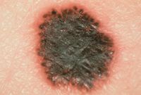
New Orleans — Several benign lesions imitate melanoma in their clinical and/or pathological presentation, scaring patients and leading to more aggressive treatment than necessary. All this can be avoided, according to Zsolt Argenyi, M.D.

"When dealing with pigmented lesions, the classical benign and malignant ones are easy to diagnose," he says. "In between is a gray zone with some unusual benign lesions."
Dr. Argenyi, director of the Pigmented Lesion Clinic and the Dermatopathology Division at the University of Washington Medical Center in Seattle, defines unusual as: appearing in a rapid fashion, having an irregular appearance or occurring in an atypical age group.

Fortunately, the condition has a characteristic appearance (a starburst-like pigment network with peripheral globules) via dermoscopy. Papules are usually less that 0.6 cm, sharply circumscribed, and symmetric. Key histologic features: symmetric, plaque-like growth pattern; sharp demarcation; and heavily pigmented spindle cells.

Deep penetrating nevus Presenting on the face of children and young adults, these small dome-shaped papules often have a dark punctum within the lesion, leading to suspicions that there have been malignant changes in a preexisting mole.
"Dermatologists have to be familiar with the nevi's histological picture and know the general criteria for malignancy to diagnose it correctly," Dr. Argenyi says.
Key histological features: wedge-shaped growth, vertical fascicles and nests, growth follows adnexal and neurovascular structures, and epithelioid and spindle cells with variable pigmentation but without significant mitotic activity.
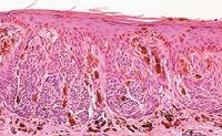
Ink spot lentigo These dark nevi have irregular, spider-like extensions at their borders, leading to frequent misdiagnosis as melanoma. A characteristic dermoscopic image helps differentiate it from malignant melanoma in situ. Histopathologic features: club-shaped elongation of rete ridges, prominent intraepidermal melanin with accentuation at the tips and bridging of the rete, transepidermal melanin elimination, no significant melanocytic increase and variably increased dermal melanophages.
Persistent melanocytic nevus Dermatologists encounter these nevi on a daily basis. What may not be apparent is that moles have been previously - and incompletely - removed. They recur with an atypical appearance and worrisome pathologic features. Reviewing the previous biopsy report is mandatory for correct diagnosis. To rule out melanoma, look for asymmetry, patchy lymphocytic infiltrate imitating focal regression, variable cytologic atypia, pagetoid spread and poor circumspection.
Congenital nevus with proliferative nodules "Babies are sometimes born with very large, heavily pigmented moles," Dr. Argenyi says. "The majority of these lesions follow a benign course, but a raised nodule can develop within the tumor, scaring parents and physicians alike. It's very important to get the right diagnosis to avoid unnecessary and extensive surgery."


