- Acne
- Actinic Keratosis
- Aesthetics
- Alopecia
- Atopic Dermatitis
- Buy-and-Bill
- COVID-19
- Case-Based Roundtable
- Chronic Hand Eczema
- Chronic Spontaneous Urticaria
- Drug Watch
- Eczema
- General Dermatology
- Hidradenitis Suppurativa
- Melasma
- NP and PA
- Pediatric Dermatology
- Pigmentary Disorders
- Practice Management
- Precision Medicine and Biologics
- Prurigo Nodularis
- Psoriasis
- Psoriatic Arthritis
- Rare Disease
- Rosacea
- Skin Cancer
- Vitiligo
- Wound Care
Publication
Article
Dermatology Times
Fast, effective tool to diagnose nail fungus
Author(s):
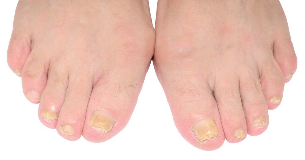
For the last several decades, global incidence rates of onychomycosis have risen. This method may be an affordable and effective tool physicians can use to improve diagnosis quickly.
For the last several decades, global incidence rates of onychomycosis have risen. This method may be an affordable and effective tool physicians can use to improve diagnosis quickly. (©ntstudio/Shutterstock.com)

For the last several decades, global incidence rates of onychomycosis have risen, but there still is no method to quickly and accurately identify which organism causes infection.
As a chronic condition, onychomycosis lingers. Without treatment, it can spread and destroy the nail. According to a new study, attenuated total reflectance Fourier transform infrared spectroscopy (ATR-FTIR) could be the fast, affordable, effective tool that improves diagnosis.
Based on the study published in the British Journal of Dermatology, the condition has been increasing in prevalence in relation to its most significant risk factors, including diabetes mellitus and occlusive footwear.1 This higher incidence makes more efficient identification of the causes behind onychomycosis a priority, particularly because the condition can present similarly to other onychopathies, such as nail psoriasis.
ATR-FTIR is showing promise in meeting this need, said study author Sander de Bruye, M.D., a microbiology, immunology, and laboratory medicine researcher at Ghent University Hospital in Belgium. This study is the first to show FTIR spectra differences between infected and uninfected nails.
“Recently, the field of FTIR spectroscopy has expanded into new and exciting clinical applications due to significant improvements in instrumentation and the development of new techniques,” Dr. de Bruye said. “In our opinion, infrared spectroscopy fits all the characteristics for the development of a new tool to diagnose onychomycosis.”
ATR-FTIR is an absorptive method that uses radiation beams to analyze and identify biological samples. Each fungal cell wall, mainly composed of glycoproteins and polysaccharides, is well-suited for this type of classification because each has a unique structure and biosynthesis. It can also recognize minor cell wall components that vary among fungi species.
This analysis surpasses current methods, including potassium hydroxide preparation, histopathological examination with periodic acid-Schiff stain, culture, and polymerase chain reaction (PCR) assays. These tactics are slower, more complex, lack sensitivity, provide false negatives, and they require trained personnel, the researchers reported.
By contrast, Dr. de Bruye said, infrared spectroscopy is fast, simple, non-destructive, and portable for in-field work. It also requires a minimal level of expertise to conduct, and it has the relatively low set-up cost of the one-time expense for the ATR-FTIR spectrometer.
To test ATR-FTIR efficacy, researchers created ex vivo and in vivo models. The ex vivo model included 158 distal fingernails and 159 toenails from 73 adults without suspected onychomycosis, and the in vivo model included distal toenails from 19 patients with possible onychomycosis. Investigators watched exvivo samples for growth over two weeks and in vivo samples for four weeks. Ex vivo nails were examined in tact, and in vivo samples were powdered to ensure homogeneity because macroscopic changes, including nail thickening or irregular surfaces, can augment spectral noise.
With the ex vivo nails, researchers conducted four classification models, differentiating between uninfected and infected nails, various dermatophytes species causing infection, as well as nails infected with non-dermatophytes and yeasts. Using dendograms, they investigated whether ATR-FTIR could be a more accessible diagnostic option.
They analyzed the spectral differences of 32 fungal microorganisms, including 20 dermatophytes (1692-1606 and 1044-1004 cm-1), 8 non-dermatophytes (973-937 cm-1), and 4 yeasts (973-937 cm-1). Using ex vivo nails, results showed prominent differences from 1200-900 cm-1 with accurate classification rates of dermatophytes, non-dermatophytes, and yeast of 91%, 97.7%, and 98.6%, respectively.
From the 19 nails in the in vivo model, 12 samples were positive for infection. Seven were identified as T. rubrum, and two were identified as Candida parapsilosis. Investigators found distinct spectral differences in the 1043-950 cm-1 region, as well as the 3000-2811 cm-1, and the 1676-1553 cm-1.
The ex vivo model revealed the carbohydrate region is the most efficient in pinpointing the discriminating features of a fungus and that FTIR spectroscopic examination of polysaccharide content could be a reliable method for detecting and differentiating which organism prompts onychomycosis.
“These results show that FTIR spectroscopic examination of the polysaccharide content could be a reliable technique for the detection and differentiation of causative organisms of onychomycosis,” Dr. de Bruye said.
In vivo model results confirmed those findings.
In addition to identifying spectral differences, Dr. de Bruye said, ATR-FTIR offers several clinical benefits. There is current debate about whether to conduct confirmatory testing before launching treatment due to the cost and time-consuming nature of the testing. ATR-FTIR could significantly reduce delays in diagnosis and treatment, he said, because it does not require any reagents or extensive sample handlings. The information gathered could also be used for providing a posteriori diagnostic analysis for other applications, including psoriasis and diabetes mellitus diagnoses. Different biochem ical fingerprints of psoriatic and onychomycotic nails could further enhance diagnostic tools, as well.
The researchers did caution, however, that while false-negative or false-positive are unlikely, ATR-FTIR might also pick up spectral characteristics from contaminating bacteria, such as green nail syndrome. In those situations, a fungal culture might still be necessary for making a correct identification and determining a proper treatment plan.
Ultimately, Dr. de Bruye said, ATR-FTIR holds a great deal of promise for improving how dermatologists approach treating onychomycosis in patients. But, more work is required.
“Based on our findings, it can be stated that ATR-FTIR spectroscopy could be a promising technique for the diagnosis of onychomycosis,” he said. “However, large-scale investigations on clinical onychomycosis diagnostics are necessary to determine and validate its definite position compared to currently employed techniques.”
Reference:
1. Boelens J, De Bruye S, Delanghe J, Hayette MP, Speeckaert M, Speeckaert R, Infrared spectroscopy as a novel tool to diagnose onychomycosis, BritishJournal of Dermatology (2018), doi: 10.1111/bjd.17199.

Newsletter
Like what you’re reading? Subscribe to Dermatology Times for weekly updates on therapies, innovations, and real-world practice tips.


