
- Acne
- Actinic Keratosis
- Aesthetics
- Alopecia
- Atopic Dermatitis
- Buy-and-Bill
- COVID-19
- Case-Based Roundtable
- Chronic Hand Eczema
- Chronic Spontaneous Urticaria
- Drug Watch
- Eczema
- General Dermatology
- Hidradenitis Suppurativa
- Melasma
- NP and PA
- Pediatric Dermatology
- Pigmentary Disorders
- Practice Management
- Precision Medicine and Biologics
- Prurigo Nodularis
- Psoriasis
- Psoriatic Arthritis
- Rare Disease
- Rosacea
- Skin Cancer
- Vitiligo
- Wound Care
Publication
Article
Dermatology Times
The Power of Mohs Surgery for Dermatofibrosarcoma Protuberans
Author(s):
Key Takeaways
- DFSP is a rare fibroblastic tumor with high recurrence rates, often misdiagnosed due to its resemblance to other skin conditions.
- Mohs micrographic surgery is the gold standard for DFSP management, offering 360° margin evaluation and tissue conservation.
A 30-year-old patient with dermatofibrosarcoma protuberans faced a recurrence after initial surgery. Mohs micrographic surgery was used for precise excision, reducing recurrence risk and improving outcomes.
“We need to get an MRI,” we told the 30-year-old patient with the help of a French interpreter. She had migrated from a West African country and was working locally in the city. She had first noticed a large, deeply invasive mass growing slowly on her left cheek over months and sought help from a hospital surgical center in the United States. The mass was excised in the operating room and closed with an advancement flap. A few days later, the pathology report showed positive margins of a dermatofibrosarcoma protuberans (DFSP). Despite the pathology report showing positive margins, she was never referred for further treatment.


Image Credit: © steheap - stock.adobe.com
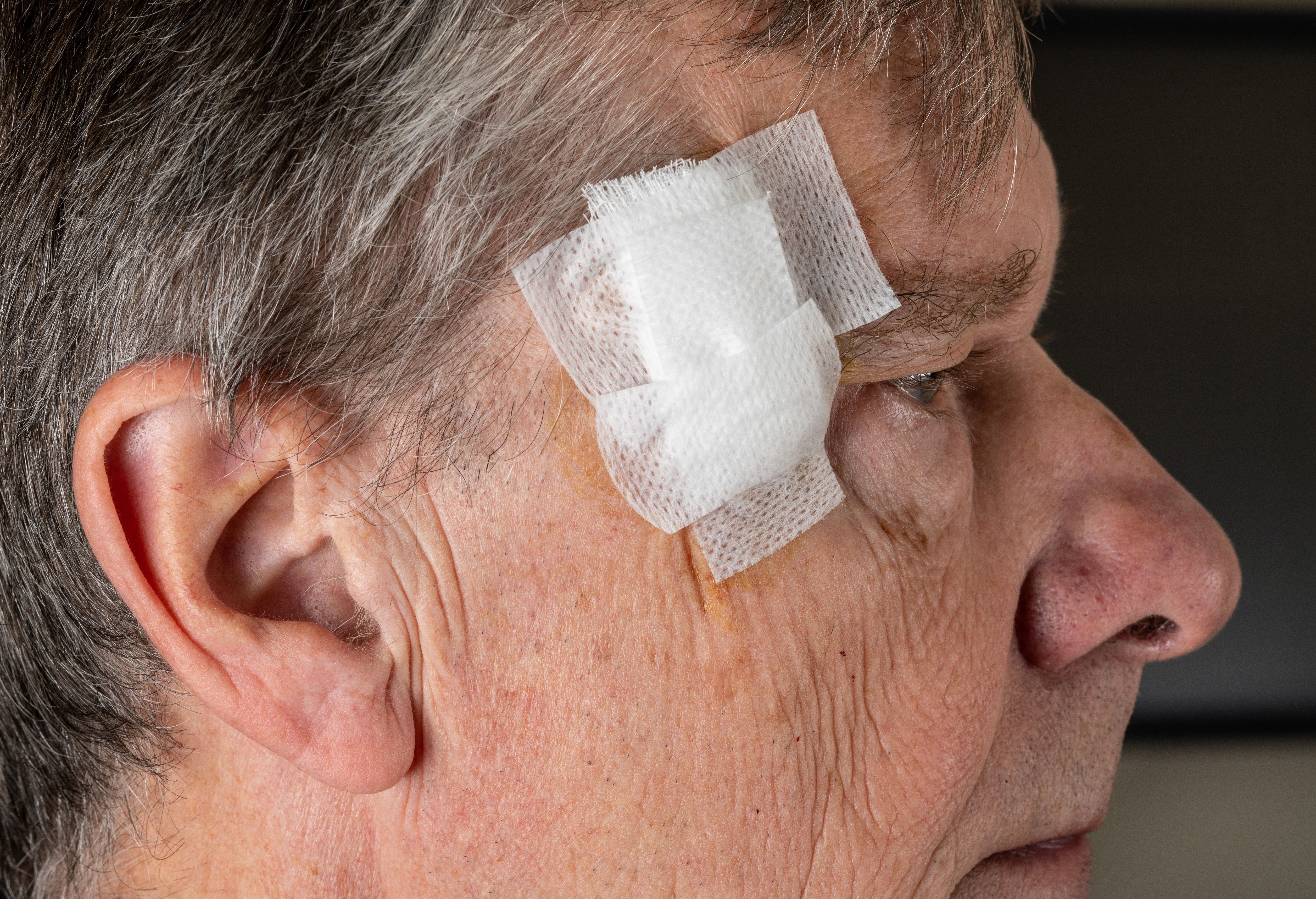
Over the next year, the mass regrew in size and slowly began to project off her cheek. She was then referred to our hospital for further evaluation and treatment. I saw her for a Mohs micrographic surgery consultation and learned about her story, the journey and pain she had been through. With a French interpreter, she spoke about her family and the social challenges she was facing at home.
On exam, the firm mass appeared fixed to the underlying structures and had ill-defined borders. We decided to order an MRI of the face to further delineate the depth of the tumor. The MRI showed that the tumor was extending into the deep soft tissue of the cheek. After discussion at the tumor board, we decided to coordinate her surgery with the Mohs; ear, nose, and throat (ENT); and plastic surgery departments. The patient’s tumor was cleared and repaired with a flap and split-thickness skin graft.
Both slow-growing and rare, DFSP has a high rate of recurrence if not completely excised. It grows in the deep dermis and subcutaneous fat, sometimes with extension to the fascia. Mohs has become one of the gold standards for surgical treatment given the tumor’s high recurrence rate and deep soft tissue infiltration. Mohs can perform a 360° margin evaluation and conserve tissue through controlled excision prior to wound closure. In this article, we explore why Mohs is an excellent treatment for DFSP.
Clinical Features of DFSP
DFSP is uncommon and represents approximately 1% of soft tissue sarcomas.1 This fibroblastic tumor often appears on the extremities and trunk but may also appear on the head and neck regions.2 It often appears as a slow-growing plaque or nodule that resembles a keloid, hypertrophic scar, cyst, or dermatofibroma. Because of DFSP’s gradual growth, there may be a delay in diagnosis before a biopsy is done in clinic. There is a median delay of 3 to 5 years for diagnosis.3 Dermatologists should be concerned about a possible DFSP if there is growth or an increasing nodular protuberant area of a lesion. Unlike other nonmelanoma skin cancers, DFSP is often diagnosed between the 2nd and 4th decades of life.2,4 Study data show that patients of African descent have a 2-fold higher rate of incidence than White patients.4 Also, the risk of DFSP metastasis is higher for tumors that are 3 cm or larger, particularly in the head, neck, and genital areas. Older, male patients who are Black have a worse prognosis.2,4 Larger tumor size and patients aged 60 and older are key prognostic factors for survival.
Histopathological Features of DFSP
DFSP is described as having whorled and monotonous spindle cells with a storiform pattern.1,5 They have positive CD34 expression and the t(17;22)(q22;q13) chromosomal translocation, generating the fusion protein COL1A1-PDGFB.1,5 There are variants of this tumor including the Bednar tumor, which is a DFSP with melanin pigment and can present as a bruise on the skin.6 An aggressive variant of a DFSP is when there is fibrosarcomatous transformation.7 Given its higher risk of metastasis, it usually requires more aggressive treatment with wider excision margins or adjuvant radiation.
Preoperative Assessment for DFSP
The day our patient came to see us, we discussed the pathology with her during her Mohs evaluation. Given her previous extensive surgery, we had enough of a tissue sample that was representative of the true pathology of the tumor. Sometimes biopsies can be too shallow to provide enough information to rule out a DFSP vs fibrosarcoma. It is important to have histopathologic confirmation with a core or incisional biopsy if the diagnosis is not clear. Pathologists can also use immunohistochemical stains such as factor XIIIa, S100, and CD34 to help differentiate DFSP from other types of tumors.8
Next, imaging can be helpful for evaluating if there is tumor invasion into underlying structures such as muscle or bone. MRI can help distinguish the depth of involvement for head and neck DFSPs. Depending on the size and depth of the tumor, it may be important to talk to patients and coordinating teams about the plan for immunotherapy vs surgery with various reconstruction options. We in the Mohs department have found it invaluable to collaborate with the ENT, radiation oncology, orthopedics, and hematology/oncology departments for complex cases at both our sarcoma and head and neck tumor boards.
Surgical Management of DFSP: Mohs Surgery
In the past, the standard treatment of DFSP was with a wide local excision (WLE) surgical margin of 2 to 4 cm.9,10 Despite taking these large margins, recurrence rates were still high at about 10% to 60% because there is often subclinical extension of the tumor. Recently, Mohs has emerged as the gold standard for surgical management of DFSP. Multiple studies have compared the recurrence rates and long-term outcomes for Mohs vs WLE. Lowe et al showed that 30.8% of patients with DFSP who had a WLE had a recurrence compared with the 3% of patients who had Mohs who had recurrence. Survival without recurrence was also higher for Mohs patients.10
Beyond decreased recurrence rates, Mohs is better for DFSP management because it uses intraoperative analysis with frozen sections.10-12 This confirms complete tumor clearance prior to surgical repair. Mohs also is used to conserve tissue in cosmetically and functionally sensitive areas such as the face and scalp, which can save patients with DFSP from a more extensive excision. Most importantly, Mohs can detect subclinical extension of the tumor at the cellular level with fresh frozen slides. Standard pathology uses serial sectioning and does not do a 360° evaluation of the surgical margin. Mohs helps to decrease the likelihood of missing foci of discontinuous tumor.


Postoperative Management and Surveillance
Patients with DFSP may end up having a very extensive Mohs surgery where the defect is larger than expected at initial evaluation. This can lead to longer healing times for patients. For these patients, we usually follow up with them post surgery at 1 to 2 weeks and then 1-month intervals to monitor healing. We make sure that patients have a scheduled follow-up with a dermatologist for a full skin exam every 6 months for at least 5 years to monitor for any recurrences. Patients can also be considered for molecular genetic testing if they have an inoperable case needing systemic therapy.13 Imatinib is a tyrosine kinase inhibitor that works best in patients with a DFSP with COL1A1-PDGFB fusion. We have had patients with inoperable, recurrent, or metastatic DFSP benefit from imatinib.
Conclusion
A rare and complex tumor, DFSP can be locally destructive and exist for years undetected or misdiagnosed. Mohs surgery has become the gold standard for DFSP management. It improves outcomes for function and reduces local recurrences. For special anatomic locations, Mohs is especially preferred over WLE as it is a precise surgery that can also spare more viable tissue. As our understanding of DFSP pathogenesis and prognosis expands, Mohs surgery continues to have a significant role in cutaneous oncology. Mohs has the power to improve survival and reduce the risk of recurrence in complex cases.
Nicole A. Negbenebor, MD, FAAD, is a Mohs micrographic surgery and cutaneous oncology clinical assistant professor and director of the Skin of Color Clinic in the Department of Dermatology at the University of Iowa in Iowa City.
References
- Menon G, Brooks J, Ramsey ML. Dermatofibrosarcoma protuberans. In: StatPearls [Internet]. StatPearls Publishing; 2025-. Updated April 18, 2024. Accessed February 17, 2025. https://www.ncbi.nlm.nih.gov/books/NBK513305/
- Kreicher KL, Kurlander DE, Gittleman HR, Barnholtz-Sloan JS, Bordeaux JS. Incidence and survival of primary dermatofibrosarcoma protuberans in the United States. Dermatol Surg. 2016;42(suppl 1):S24-31. doi:10.1097/DSS.0000000000000300
- David MP, Funderburg A, Selig JP, et al. Perspectives of patients with dermatofibrosarcoma protuberans on diagnostic delays, surgical outcomes, and nonprotuberance. JAMA Netw Open. 2019;2(8):e1910413. doi:10.1001/jamanetworkopen.2019.10413
- Maghfour J, Genelin X, Olson J, Wang A, Schultz L, Blalock TW. The epidemiology of dermatofibrosarcoma protuberans incidence, metastasis, and death among various population groups: a Surveillance, Epidemiology, and End Results database analysis. J Am Acad Dermatol. 2024;91(5):826-833. doi:10.1016/j.jaad.2024.05.088
- Trinidad CM, Wangsiricharoen S, Prieto VG, Aung PP. Rare variants of dermatofibrosarcoma protuberans: clinical, histologic, and molecular features and diagnostic pitfalls. Dermatopathology (Basel). 2023;10(1):54-62. doi:10.3390/dermatopathology10010008
- Almeida FT, Carvalho SD, Pereira T, Brito C. When a bruise gets important: Bednar tumour. BMJ Case Rep. 2019;12(2):e228446. doi:10.1136/bcr-2018-228446
- Liang CA, Jambusaria-Pahlajani A, Karia PS, Elenitsas R, Zhang PD, Schmults CD. A systematic review of outcome data for dermatofibrosarcoma protuberans with and without fibrosarcomatous change. J Am Acad Dermatol. 2014;71(4):781-786. doi:10.1016/j.jaad.2014.03.018
- Hardy CSC, Razavi A, Nunez N, et al. (2024). Immunohistochemical profiles of dermatofibroma and dermatofibrosarcoma protuberans: a scoping review. medRxiv. Preprint posted online October 28, 2024. doi:10.1101/2024.10.23.24316006
- Snow SN, Gordon EM, Larson PO, BagheriM M, Bentz ML, Sable DB. Dermatofibrosarcoma protuberans: a report on 29 patients treated by Mohs micrographic surgery with long‐term follow‐up and review of the literature. Cancer. 2004;101(1):28-38. doi:10.1002/cncr.20316
- Lowe GC, Onajin O, Baum CL, et al. A comparison of Mohs micrographic surgery and wide local excision for treatment of dermatofibrosarcoma protuberans with long-term follow-up: the Mayo Clinic experience. Dermatol Surg. 2017;43(1):98-106. doi:10.1097/DSS.0000000000000910
- Siddiqui FS, Sathe NC, Leavitt A. Mohs micrographic surgery evaluation and treatment of dermatofibrosarcoma protuberans. In: StatPearls [Internet]. StatPearls Publishing; 2025-.
- Martin ECS, Vyas KS, Batbold S, Erwin PJ, Brewer JD. Dermatofibrosarcoma protuberans recurrence after wide local excision versus Mohs micrographic surgery: a systematic review and meta-analysis. Dermatol Surg. 2022;48(5):479-485. doi:10.1097/DSS.0000000000003411
- Wright TI, Petersen JE. Treatment of recurrent dermatofibrosarcoma protuberans with imatinib mesylate, followed by Mohs micrographic surgery. Dermatol Surg. 2007;33(6):741-744. doi:10.1111/j.1524-4725.2007.33154.x


Newsletter
Like what you’re reading? Subscribe to Dermatology Times for weekly updates on therapies, innovations, and real-world practice tips.


