- Acne
- Actinic Keratosis
- Aesthetics
- Alopecia
- Atopic Dermatitis
- Buy-and-Bill
- COVID-19
- Case-Based Roundtable
- Chronic Hand Eczema
- Chronic Spontaneous Urticaria
- Drug Watch
- Eczema
- General Dermatology
- Hidradenitis Suppurativa
- Melasma
- NP and PA
- Pediatric Dermatology
- Pigmentary Disorders
- Practice Management
- Precision Medicine and Biologics
- Prurigo Nodularis
- Psoriasis
- Psoriatic Arthritis
- Rare Disease
- Rosacea
- Skin Cancer
- Vitiligo
- Wound Care
Publication
Article
Dermatology Times
Pemphigus: Updated Review and Emerging Therapies
Author(s):
Pemphigus is defined as a group of autoimmune, blistering disorders caused by circulating autoantibodies that bind to desmogleins, proteins that are vital to epidermal intercellular adhesion.
Overview of Pemphigus
Pemphigus is defined as a group of autoimmune, blistering disorders caused by circulating autoantibodies that bind to desmogleins (Dsg), which are proteins that are vital to epidermal intercellular adhesion.1 Binding of these autoantibodies to their targets causes acantholysis (or loss of keratinocyte to keratinocyte adhesion), which ultimately results in the formation of intraepithelial blisters in skin and mucous membranes.1 Unfortunately, pemphigus disorders can potentially be life-threatening and have a devastating impact on quality of life.
Pemphigus vulgaris and pemphigus foliaceus, which comprise 90–95% of pemphigus diagnoses, will be described here.2,3 Other types of pemphigus include Immunoglobulin A (IgA) pemphigus, Paraneoplastic pemphigus, Drug-induced pemphigus, Pemphigus vegetans, Pemphigus erythematosus (Senear-Usher syndrome), Pemphigus herpetiformis, Atypical pemphigus, and Neonatal pemphigus.2,4,5
Pemphigus diseases are different from pemphigoid diseases. Pemphigoid diseases are the other main group of autoimmune blistering disorders and are characterized by autoantibodies against structural proteins of the dermal–epidermal junction.6 Pemphigus diseases demonstrate a positive Nikolsky’s sign (extension of a blister when pressure is applied to the side of the blister), whereas Pemphigoid diseases do not.1
Diagnosis of pemphigus disorders is often aided by clinical presentation, direct immunofluorescence microscopy of a perilesional biopsy, serology, and histopathology of a lesional biopsy.7
Systemic corticosteroids represent the first-line treatment in all forms of pemphigus.7 Adjuvant medications include azathioprine and mycophenolate compounds.7 Rituximab, an anti-CD20 monoclonal antibody that induces B-cell depletion, has been shown to improve patient survival.8 Currently, rituximab in conjunction with corticosteroids is first-line for treatment of moderate to severe pemphigus, thus allowing for a major corticosteroid-sparing effect and decreasing the risk of steroid-associated adverse events.7
Pemphigus vulgaris (PV)
PV, the most common type of pemphigus, causes blisters on cutaneous and mucosal surfaces.
The mean age of onset is 50 to 60 years of age, and it affects both sexes equally.9 The reported incidence is between 0.1 and 0.5 per 100,000 people per year, but can be higher in certain groups, such as Ashkenazi Jews.9
In terms of etiology, genetics are thought to confer susceptibility to the disease. Environmental factors, including stress, diet, and infection, may trigger immune dysregulation, leading to flares.9 Patients with PV have autoantibodies against Dsg3 or both Dsg3 and Dsg1.9
In 80% of PV cases, patients initially present with intraoral blisters that frequently rupture, resulting in painful erosions.9 In 75% of PV cases, cutaneous lesions arise after the first oral blisters.9 Physical exam may reveal vesicles, erosions, or bullae on erythematous or normal-appearing skin.9
PV in a 33-year-old man.
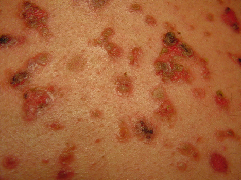
Diagnostic clues favoring PV include mucosal lesions, positive Nikolsky’s sign, positive direct immunofluorescence microscopy results (i.e., intercellular deposits of IgG, complement component 3, or both), and serum antibodies against Dsg3.7
As in all forms of pemphigus, corticosteroids are first-line in the treatment of PV.7 There is an accepted algorithm for the management of PV developed by international experts, which resulted from a Delphi consensus based on the European Dermatology Forum and the European Academy of Dermatology and Venereology guidelines.7
Despite advances in treatment, patients with PV should be monitored closely. Patients can experience relapses and complications, such as infection. In fact, septicemia is the leading cause of death in PV, indicating that there is still much progress to be made with PV management.9
Pemphigus foliaceus (PF)
Unlike PV, PF is characterized by skin involvement only, with the sparing of mucosal surfaces.
PF is less common than PV. The mean age of onset is 50 to 60 years of age, and it affects both sexes equally.10Endemic pemphigus foliaceus, which is also known as Fogo selvagem, is common in Brazil and other South American countries.10, 11
Regarding etiology, genetics are thought to confer susceptibility to the disease, while environmental factors, such as the use of certain drugs, act as triggers. In particular, penicillamine and captopril, which contain sulfhydryl groups that are speculated to interact with the sulfhydryl groups in Dsg1 and Dsg3, are associated with drug-induced PF.10
Most patients with PF are not severely ill.10 Patients may suffer from burning, pruritus, and pain.10 The onset is usually subtle with a few scattered lesions involving the seborrheic areas (i.e., the scalp, face, chest, and upper back).10 The lesions are characterized by erythema, “puff pastry-like” scaling, and crusting.3 Intact blisters are rarely seen because they are superficial and rupture easily.10 PF may stay localized for years or quickly progress to generalized involvement, leading to exfoliative erythroderma.10
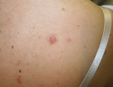
PF in a 61-year-old woman.
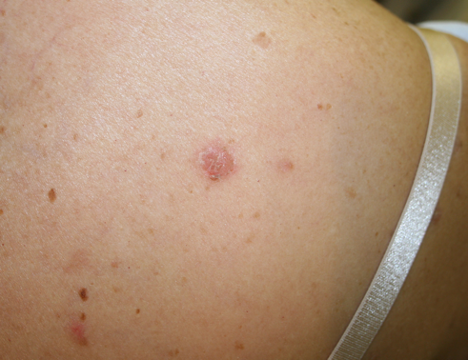
The diagnosis of PF is supported by crusted erosions, lack of mucosal lesions, positive Nikolsky’s sign, positive direct immunofluorescence microscopy results (i.e., intercellular deposits of IgG, complement component 3, or both), and serum antibodies against Dsg1.7
As in all forms of pemphigus, systemic corticosteroids are first-line in the treatment of PF.7 The treatment algorithm for PF is similar to that of PV.7 However, in localized forms of PF with a limited number of lesions, topical or intralesional corticosteroids can be used.12
Prior to corticosteroid therapy, PF was fatal in approximately 60% of patients.10 Fortunately, steroids and other immunosuppressive therapies have greatly reduced mortality.10 In terms of prognosis, PF is a benign disease that responds well to treatment and may remit.10
Pemphigus: Emerging Therapies
There are many new treatments currently being tested for pemphigus.3,8,13,14,15 Second and third generation anti-CD20 monoclonal antibodies, such as veltuzumab, ocrelizumab, and obinutuzumab, have higher binding affinities and induce a more robust B-cell depletion compared to rituximab.3,13 The development of these next generation anti-CD20 monoclonal antibodies could lead to major progress in the treatment of pemphigus. Bruton tyrosine kinase (BTK) inhibitors, which downregulate several B-cell functions, and efgartigimod, an engineered Fc fragment derived from human IgG1, are also being studied for use in pemphigus treatment.13,14,15 Recently, researchers have engineered chimeric autoantibody receptor (CAAR)-T cells that recognize Dsg domains to selectively target autoreactive B-cells, which could become a precise therapy for pemphigus in the future.3,8 Other novel, potential therapies include cytokine inhibitors, anti-CD25 monoclonal antibodies, autologous hematopoietic stem cell transplantation, and polyclonal regulatory T cell (PolyTreg) therapy.13 Hopefully, one or more of these emerging therapies will soon be shown to be safe and effective in the treatment of pemphigus, and ultimately improve survival and quality of life for these patients.3,8,13
References
1. Schmidt E, Kasperkiewicz M, Joly P. Pemphigus. The Lancet. 2019;394(10201):882-894. doi:10.1016/S0140-6736(19)31778-7
2. Kridin K, Schmidt E. Epidemiology of Pemphigus. JID Innov. 2021;1(1):100004. doi:10.1016/j.xjidi.2021.100004
3. Lim YL, Bohelay G, Hanakawa S, Musette P, Janela B. Autoimmune Pemphigus: Latest Advances and Emerging Therapies. Front Mol Biosci. 2022;8:808536. doi:10.3389/fmolb.2021.808536
4. Di Lernia V, Casanova DM, Goldust M, Ricci C. Pemphigus Vulgaris and Bullous Pemphigoid: Update on Diagnosis and Treatment. Dermatol Pract Concept. 2020;10(3):e2020050. doi:10.5826/dpc.1003a50
5. Pemphigus: Types, Symptoms, Causes & Treatment. Cleveland Clinic. Accessed November 23, 2022. https://my.clevelandclinic.org/health/diseases/21130-pemphigus
6. Schmidt E, Zillikens D. Pemphigoid diseases. Lancet Lond Engl. 2013;381(9863):320-332. doi:10.1016/S0140-6736(12)61140-4
7. Murrell DF, Peña S, Joly P, et al. Diagnosis and management of pemphigus: Recommendations of an international panel of experts. J Am Acad Dermatol. 2020;82(3):575-585.e1. doi:10.1016/j.jaad.2018.02.021
8. Didona D, Maglie R, Eming R, Hertl M. Pemphigus: Current and Future Therapeutic Strategies. Front Immunol. 2019;10:1418. doi:10.3389/fimmu.2019.01418
9. Ingold CJ, Khan MA. Pemphigus Vulgaris. In: StatPearls. StatPearls Publishing; 2022. Accessed December 11, 2022. http://www.ncbi.nlm.nih.gov/books/NBK560860/
10. Lepe K, Yarrarapu SNS, Zito PM. Pemphigus Foliaceus. In: StatPearls. StatPearls Publishing; 2022. Accessed December 11, 2022. http://www.ncbi.nlm.nih.gov/books/NBK499829/
11. Hans-Filho G, Aoki V, Bittner NRH, Bittner GC. Fogo selvagem: endemic pemphigus foliaceus. An Bras Dermatol. 2018;93(5):638-650. doi:10.1590/abd1806-4841.20188235
12. Porro AM, Filho GH, Santi CG. Consensus on the treatment of autoimmune bullous dermatoses: pemphigus vulgaris and pemphigus foliaceus - Brazilian Society of Dermatology. An Bras Dermatol. 2019;94(2 Suppl 1):20. doi:10.1590/abd1806-4841.2019940206
13. Yuan H, Pan M, Chen H, Mao X. Immunotherapy for Pemphigus: Present and Future. Front Med. 2022;9. Accessed December 11, 2022. https://www.frontiersin.org/articles/10.3389/fmed.2022.901239
14. argenx. A Randomized, Double-Blinded, Placebo-Controlled Trial to Investigate the Efficacy, Safety, and Tolerability of Efgartigimod PH20 SC in Adult Patients With Pemphigus (Vulgaris or Foliaceus). clinicaltrials.gov; 2022. Accessed December 8, 2022. https://clinicaltrials.gov/ct2/show/NCT04598451
15. argenx. An Open-Label, Multicenter, Follow-up Trial of ARGX-113-1904 to Evaluate the Safety, Tolerability, and Efficacy of Efgartigimod PH20 SC in Patients With Pemphigus. clinicaltrials.gov; 2022. Accessed December 8, 2022. https://clinicaltrials.gov/ct2/show/NCT04598477
Newsletter
Like what you’re reading? Subscribe to Dermatology Times for weekly updates on therapies, innovations, and real-world practice tips.

