- Acne
- Actinic Keratosis
- Aesthetics
- Alopecia
- Atopic Dermatitis
- Buy-and-Bill
- COVID-19
- Case-Based Roundtable
- Chronic Hand Eczema
- Chronic Spontaneous Urticaria
- Drug Watch
- Eczema
- General Dermatology
- Hidradenitis Suppurativa
- Melasma
- NP and PA
- Pediatric Dermatology
- Pigmentary Disorders
- Practice Management
- Precision Medicine and Biologics
- Prurigo Nodularis
- Psoriasis
- Psoriatic Arthritis
- Rare Disease
- Rosacea
- Skin Cancer
- Vitiligo
- Wound Care
Publication
Article
Dermatology Times
Optimize Risk, Management of Pigmented Lesions in Pediatric Patients
Author(s):
Diagnosis, risk assessment and treatment of congenital melanocytic nevi (CMN), as well as melanoma, require specific strategies for pediatric patients.
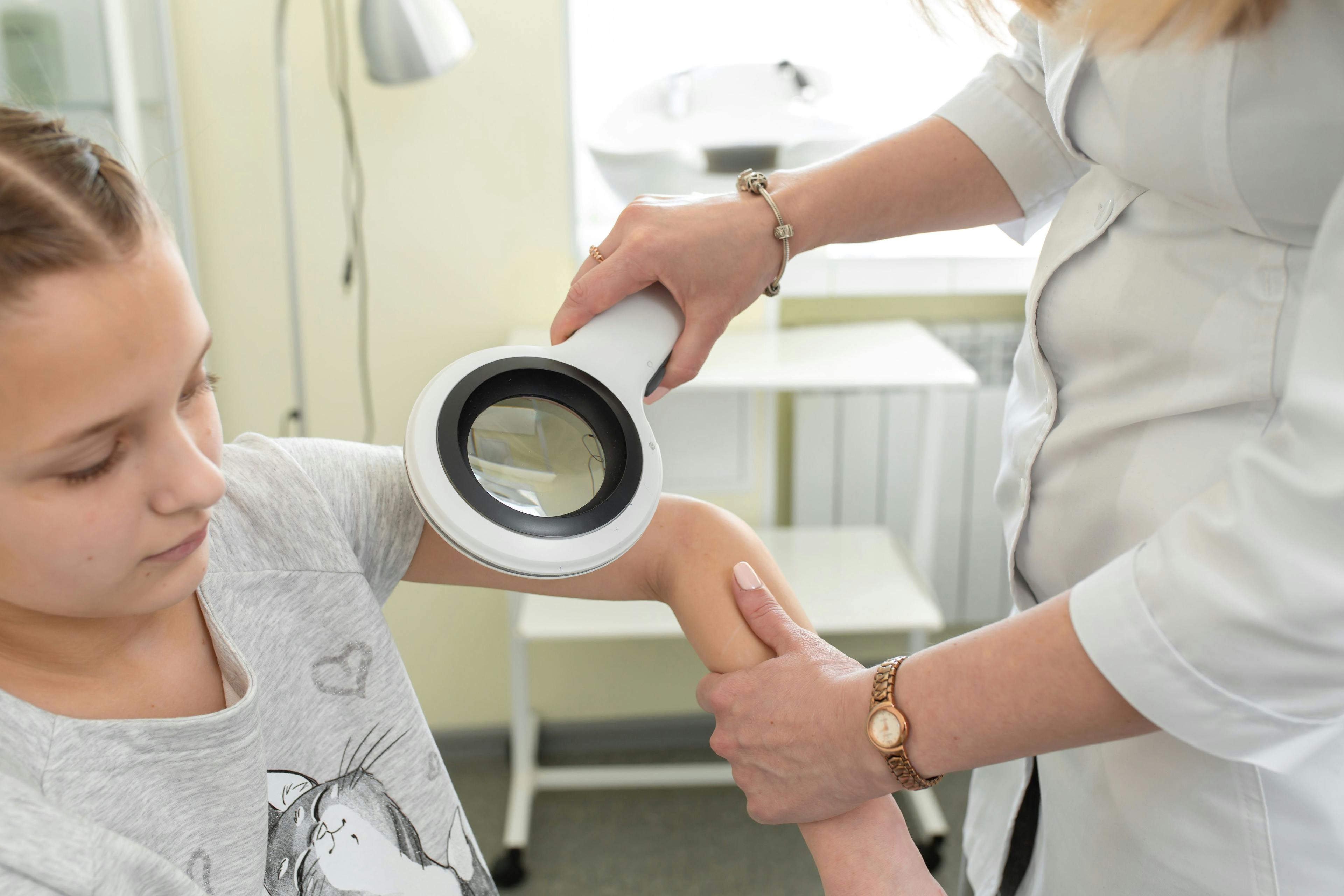
Diagnosis, risk assessment and treatment management of congenital melanocytic nevi (CMN) as well as melanoma require specific strategies for pediatric patients. Elena B. Hawryluk, MD, PhD, assistant professor of dermatology at Harvard Medical School in Boston, Massachusetts, discussed recommendations for understanding how melanoma presents in pediatric cases and how to optimize outcomes during her presentation at Maui Derm Live. The in-person dermatology continuing medical education (CME) conference in Hawaii was held concurrently with Maui Derm Connect, a virtual CME conference, January 25 to January 29, 2021.1
Assessing CMN Risks
Hawryluk first addressed CMN, noting that they increase the risk of melanoma and that this risk increases with the size of the nevus. For small (<1.5cm) and medium (1.5 to 20 cm) nevi, the risk increases by roughly 1%. For large (>20cm) and giant (>40cm) nevi, the risk is 2% for the large and 6% to 15% for the giant. In a study of CMN-associated melanomas, 78% had giant size and 95% had many satellite nevi.
She added that atypical or dysplastic nevi, which are not necessarily pre-melanoma, are an indication of increased risk of developing melanoma. Because of this, Hawryluk recommended a thorough examination along with continual surveillance.
Pediatric patients with large or giant nevi who develop melanoma have high mortality (55%) and 14% have visceral melanoma, she said. Hawryluk also noted study results showing that patients with 2 or more nevi have an increased risk of central nervous system involvement, with 21% of the patients having abnormal magnetic resonance imaging (MRI) findings.2
Is It Melanoma?
Hawryluk noted that various types of melanoma affect the pediatric population and generally present differently in pediatric versus adult patients. A 2013 study3 advised assessment of these lesion characteristics when diagnosing pediatric cases: amelanotic, bump, bleeding, color uniformity, de novo, at any diameter.
Amelanotic melanoma can often be incorrectly diagnosed as a benign lesion. Hawryluk cited a 2016 study from the Journal of the American Academy of Dermatology that found dermatologists had identified an amelanotic melanoma as a spitz nevus, hemangioma, acneiform lesion, and spitz nevus following a first impression. She shared a case from her practice involving a girl with a suspected wart that hadn’t responded to all typical treatments, to illustrate the point.
Spitzoid melanoma can harbor kinase fusions in roughly 50% of lesions and can be difficult to diagnose and distinguish from atypical Spitz tumors, in part because non-melanoma spitz lesions can also harbor kinase fusions, she added.
Optimize Workups and Management Options
If there is no suspicion of melanoma, some parents prefer simply to keep the lesions under observation. According to Hawryluk, other choices include surgical excision, although no strong evidence has been found that it reduces the risk of melanoma; curettage; dermabrasion; chemical peels; cryotherapy; laser; and electrotherapy. These choices can remove CMN cells, but Hawryluk remarked that they carry the risk of scar or disfigurement and may make it more difficult to detect melanoma.
When considering whether a child needs an MRI, she recommended that it be done to infants who had 2+ nevi, a large axial nevus, the presence of satellites, or in older patients who are exhibiting neurologic change. She also noted that having imaging from infancy to serve as a baseline can make it easier to discern changes.
For the workup of patients with CMN and melanoma suspicion, she shared two algorithms4 dependent on whether the presentation was cutaneous or a central nervous system manifestation.
For cutaneous presentations, a thorough clinical examination with photo documentation should occur with a review with 4 weeks. If the new mark is resolved or unchanged, the area should be monitored continually to ensure stability. If the mark progresses or has an abnormal presentation, an excision biopsy should occur, along with histopathology and hotspot genotyping.
If it’s malignant and confirmed by another expert, baseline blood, histology, staging, and further excision should occur as dictated. For new neurological symptoms, imaging should occur. If it’s normal or unchanged since the last imaging session, regular monitoring should continue. For abnormal results, biopsy followed by histopathology and genotyping can be used to determine if the result is malignant or nonmalignant.
Disclosure:
Hawryluk reported no relevant or financial disclosure.
References:
1.Hawryluk E. The pigmented lesion clinic: pediatric cases. Oral presentation at: MauiDerm for Dermatologists Live Dermatology CME Conference and Virtual Dermatology CME Conference held concurrently; January 25-29, 2021; Maui, Hawaii; virtual.
2.Waelchli R, et al. Classification of neurological abnormalities in children with congenital melanocytic naevus syndrome identifies magnetic resonance imaging as the best predictor of clinical outcome. Br J Dermatol. 2015. 173(3): p. 739-50.
3.Cordoro KM, Gupta D, Frieden IJ, McCalmont T, Kashani-Sabet M. Pediatric melanoma: results of a large cohort study and proposal for modified ABCD detection criteria for children. J Am Acad Dermatol. 2013;68(6):913-925. doi:10.1016/j.jaad.2012.12.953
4.Kinsler VA, O'Hare P, Bulstrode N, Calonje JE, Chong WK, Hargrave D, Jacques T, Lomas D, Sebire NJ, Slater O. Melanoma in congenital melanocytic naevi. Br J Dermatol. 2017 May;176(5):1131-1143. doi: 10.1111/bjd.15301. Epub 2017 Apr 4. PMID: 28078671; PMCID: PMC5484991.
Newsletter
Like what you’re reading? Subscribe to Dermatology Times for weekly updates on therapies, innovations, and real-world practice tips.


