- Acne
- Actinic Keratosis
- Aesthetics
- Alopecia
- Atopic Dermatitis
- Buy-and-Bill
- COVID-19
- Case-Based Roundtable
- Chronic Hand Eczema
- Chronic Spontaneous Urticaria
- Drug Watch
- Eczema
- General Dermatology
- Hidradenitis Suppurativa
- Melasma
- NP and PA
- Pediatric Dermatology
- Pigmentary Disorders
- Practice Management
- Precision Medicine and Biologics
- Prurigo Nodularis
- Psoriasis
- Psoriatic Arthritis
- Rare Disease
- Rosacea
- Skin Cancer
- Vitiligo
- Wound Care
Publication
Article
Dermatology Times
PET-CTs for melanoma may have less benefit than previously thought
Author(s):
Stage II melanoma patients don’t always benefit from staging PET-CT, according to Duke University Medical Center researchers reporting last week at American Society of Dermatologic Surgery annual meeting in Phoenix.
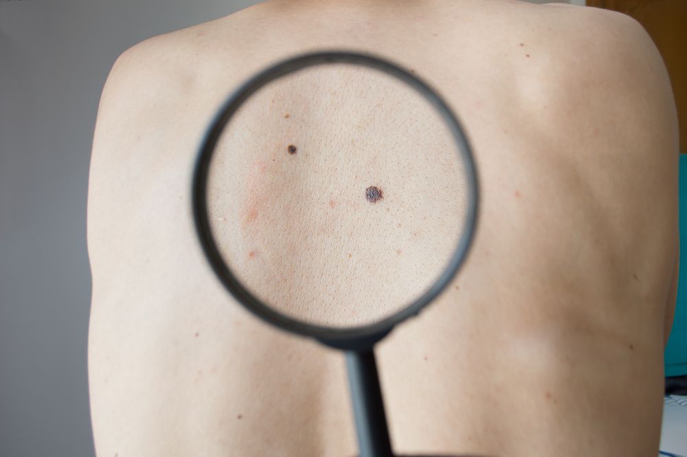
Stage II melanoma patients don’t always benefit from staging PET-CT, according to Duke University Medical Center researchers reporting last week at American Society of Dermatologic Surgery annual meeting in Phoenix. (©Cunaplus/Shutterstock.com)

PHOENIXâPET-CT is often ordered reflexively when clinicians are concerned about melanoma metastasis, but the potential for harm from false positive results is not always weighed.
A study to evaluate the usefulness of PET-CT in staging patients with stage II melanoma was conducted at Duke University Medical Center, with a retrospective review of all patients who were imaged over an 11-year period, from 2005 to 2016. The abstract was presented last week at the American Society of Dermatologic Surgery annual meeting in Phoenix.
“We frequently observed that PET-CTs performed for melanoma in our institution were of less utility than we may have hoped,” said study author Adam Brys, M.D., a second year dermatology resident at Duke Hospital with a special interest in melanoma. “Costs of healthcare modalities are also crucial to consider, as avoiding unnecessary tests allow better utilization and distribution of limited resources to those patients who truly would benefit from such tests.”
During the study period, 225 patients had a PET-CT scan within three months after their initial melanoma diagnosis, and more than half of them had it in conjunction with a sentinel lymph node biopsy, while some palliative cases chose to forego the biopsy. Out of these, 89 patients had stage II melanoma.
Metastasis was detected in only one patient, who had an ulcerated primary melanoma with a Breslow thickness of 7 mm. So the authors concluded that staging with PET-CT in patients with stage II melanoma rarely provides information that is likely to change management.
The study focused on both stage II and III melanoma, as patients with advanced stage II disease often have a better five-year survival rate than patients with less advanced stage III disease.
“For example, a very thick melanoma with no nodal metastasis has a worse prognosis than a thin melanoma with minimal, but positive, nodal involvement,” Dr. Brys said.
In a separate presentation at ASDS, the authors also presented data on patients with stage III melanoma who presented with occult positive lymph nodes that were detected only through sentinel node biopsies.
He pointed out that current guidelines, based on moderate strength data and some consensus, recommend no imaging in patients with stage II melanoma, although they state it should be considered in patients with AJCC stage IIIA melanoma and obtained in other stage III melanoma cases.
Patients with new symptoms suggestive of metastatic disease and patients with clinically detected, palpable lymph nodes should undergo imaging. But for asymptomatic patients with thin melanomas, cross sectional imaging will not provide benefits.
However, further study is warranted to identify strategies for accurately predicting which patients would be most likely to benefit from baseline staging PET-CT, and to determine more precise prognostic factors, such as gene mutations or precise thickness, that may be predictive of the utility of PET-CT.
“It should be noted that certain clinical trials may require baseline imaging. And of course, exceptions certainly exist to all guidelines and recommendations,” Dr. Brys said.
REFERENCE
Brys, A. (2018). American Society of Dermatologic Surgery, Phoenix, Arizona. October 11, 2018. Utility of Staging PET-CT in Patients with Stage II Melanoma. Poster # 2

Newsletter
Like what you’re reading? Subscribe to Dermatology Times for weekly updates on therapies, innovations, and real-world practice tips.


