- Acne
- Actinic Keratosis
- Aesthetics
- Alopecia
- Atopic Dermatitis
- Buy-and-Bill
- COVID-19
- Case-Based Roundtable
- Chronic Hand Eczema
- Chronic Spontaneous Urticaria
- Drug Watch
- Eczema
- General Dermatology
- Hidradenitis Suppurativa
- Melasma
- NP and PA
- Pediatric Dermatology
- Pigmentary Disorders
- Practice Management
- Precision Medicine and Biologics
- Prurigo Nodularis
- Psoriasis
- Psoriatic Arthritis
- Rare Disease
- Rosacea
- Skin Cancer
- Vitiligo
- Wound Care
Publication
Article
Dermatology Times
Mast cells may play key role in rosacea pathogenesis
Author(s):
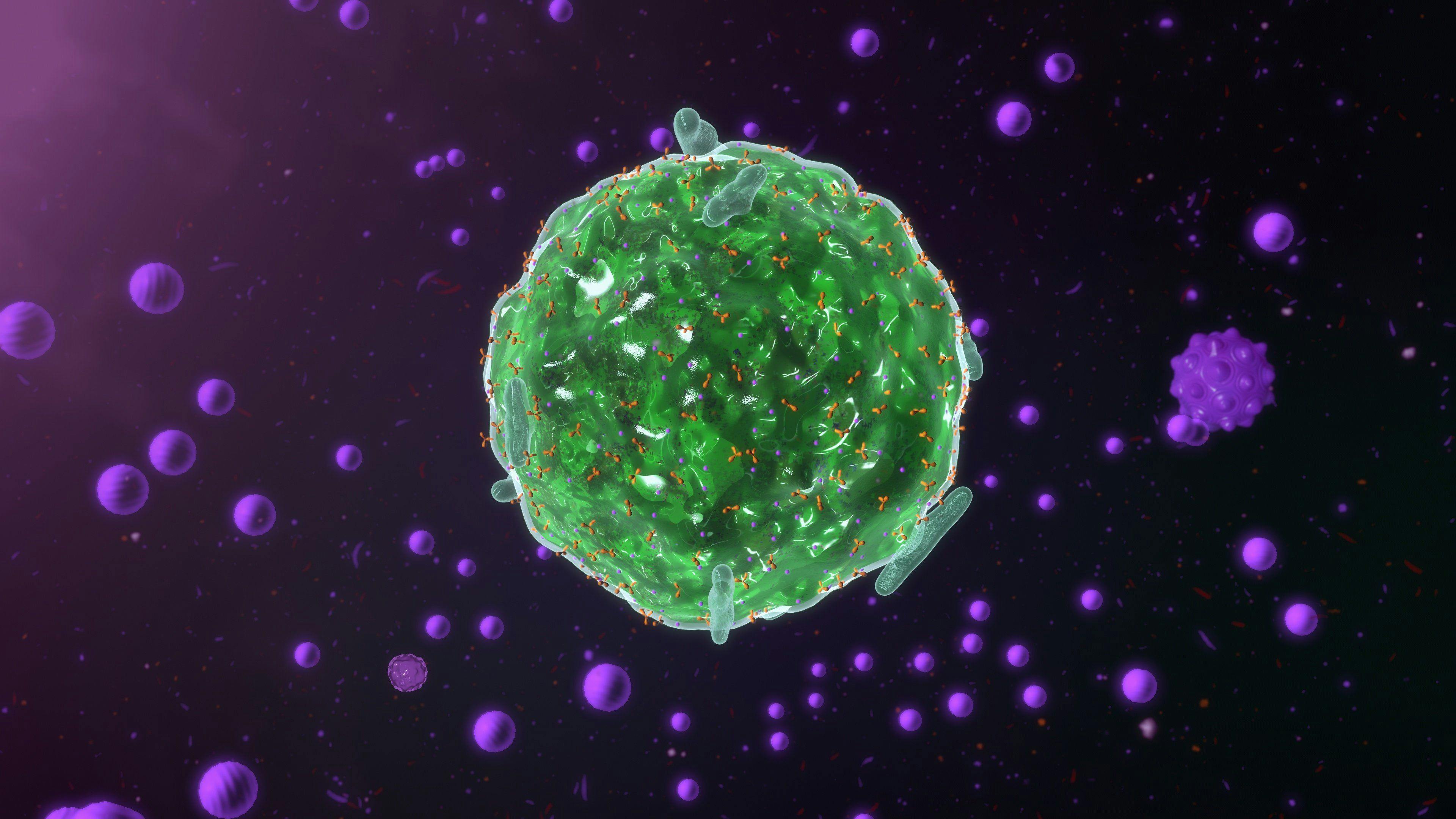
Mast cells appear to play an intricate role in the pathophysiology of rosacea and could serve as potential targets for future therapies, according to recent study findings.
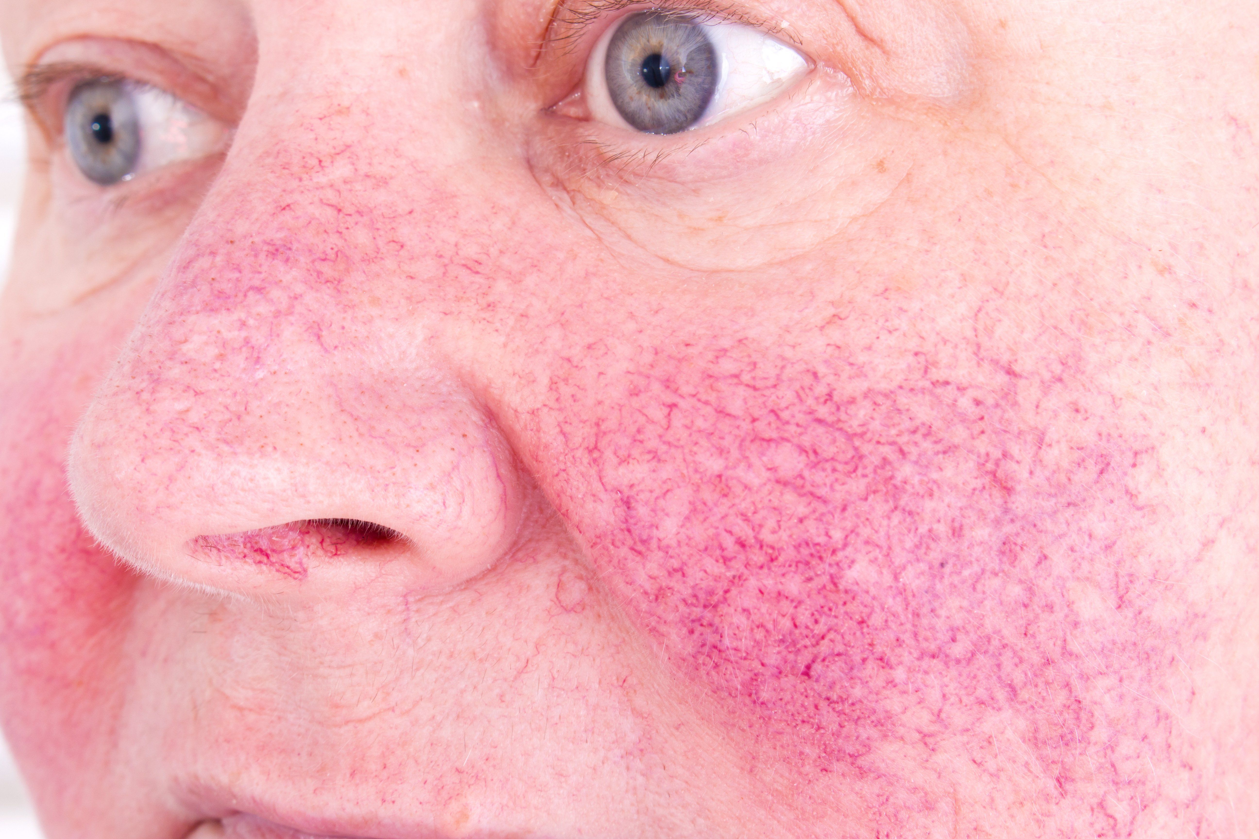
Mast cells appear to play an intricate role in the pathophysiology of rosacea and could serve as potential targets for future therapies, according to recent study findings1.
RELATED: A new target for rosacea treatment: The mast cell
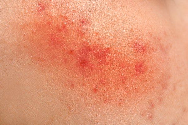
Impacting approximately 5% of the adult population worldwide, rosacea can significantly impact a patient’s quality of life and lead to self-abasement, depression, anxiety and social phobia.
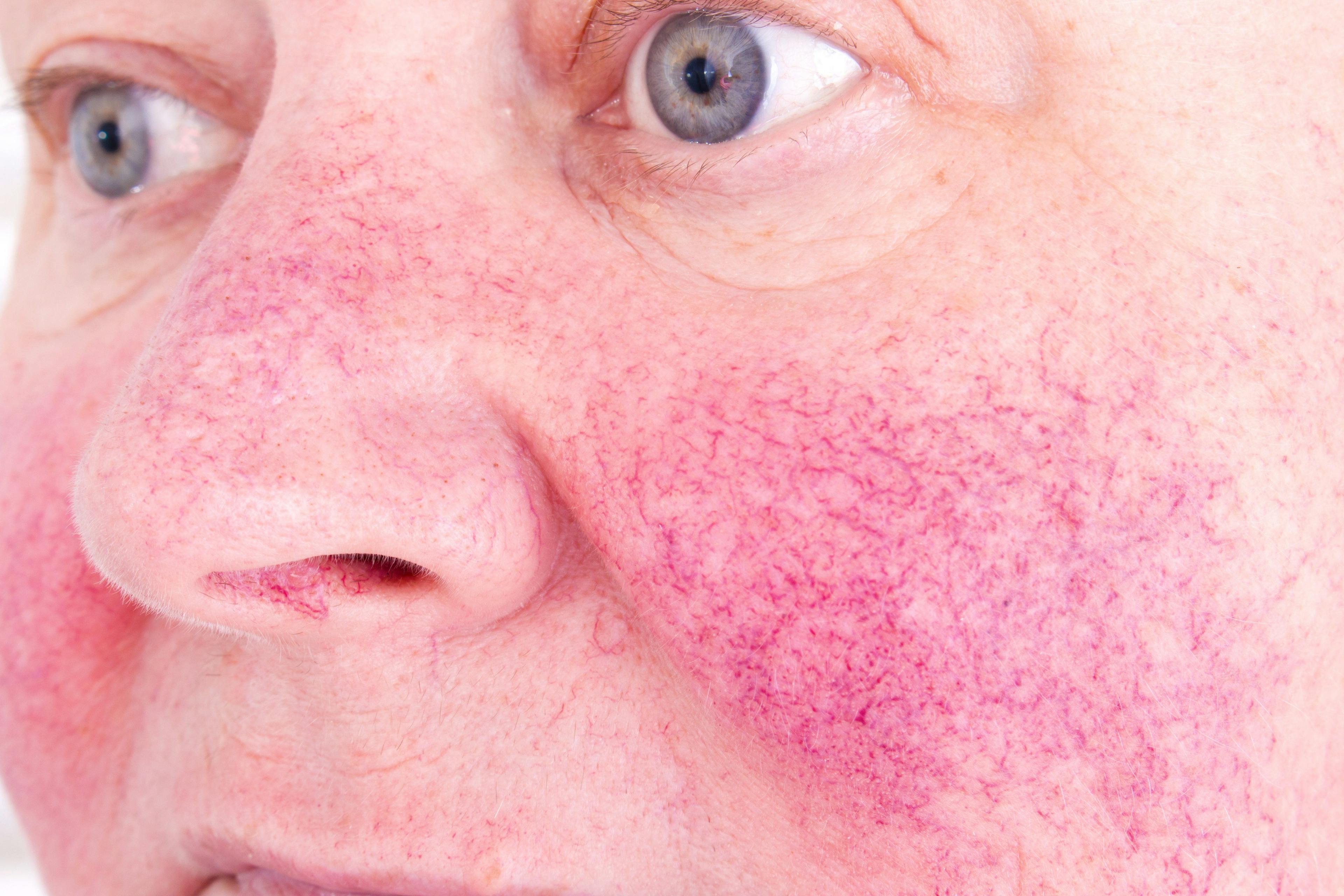
Heralded by transient/persistent erythema and inflammatory papules/pustules with or without telangiectasia, rosacea is further divided into four subtypes including erythematotelangiectatic rosacea, papulopustular rosacea, phymatous rosacea or rhinophyma, and ocular rosacea.
“Rosacea is a complex disorder caused by immune dysfunction and neurovascular dysregulation involving many types of inflammatory cells,” writes Lin Wang, M.D., and fellow colleagues, department of dermatology, West China Hospital, Sichuan University, Chengdu, China, in the paper that recently appeared in Frontiers in Medicine.
“Microbes, ultraviolet radiation, temperature, stress, and a damaged skin barrier can be triggers, stimulating an augmented innate immune response and/or neurovascular dysregulation.”
It is widely known that mast cells reside in virtually all tissues throughout the body and are particularly abundant in the skin and mucosal surfaces. As such, mast cells are one of the first cells of the immune system that interact with environmental antigens, allergens and invasive pathogens.
The differentiation, survival and migration of mast cells require a stem cell factor, which can be regulated by keratinocytes, the study authors write, and the activation and migration of mast cells play important roles in regulating homeostasis.
“Once activated, the mast cells can promote the release of different mediators and have a considerable effect on the pathophysiology of diverse inflammatory diseases,” Dr. Wang et al. write.
RELATED: Education key to optimizing care of rosacea patients
These mediators include histamine, tryptase, and chymase, which are preformed mediators stored in granules that initiate the allergic cascade. Effector molecules including platelet activating factor and prostaglandins can be synthesized upon cell stimulation the authors say, and cytokines including interleukin-1, transforming growth factor-beta, TNF-alpha, and vascular endothelial growth factor can be released, further activating the mast cells.
“These effectors can participate in numerous inflammatory responses and endothelial dysfunction in the vasculature, altering neural activity and function,” the study authors write.
The functional characterization of mast cells in rosacea is only recently emerging. One study2 showed that the number of mast cells was significantly higher in lesions than in clinically uninvolved skin in patients with papulopustular and erythematotelangiectatic rosacea, and there was a positive correlation between mast cell density and the duration of rosacea.
According to the authors, adaptive immunity along with the innate immune system might play a critical role in the pathophysiology of rosacea; past studies have shown that mast cells can heighten host defense by initiating inflammation associated with innate immune responses.
“Although there are no reports on the interaction between mast cells and other immune cells, including macrophages, neutrophils and T cells in rosacea, many studies have proven that mast cells can be activated by other immune cells or can activate neighboring cells through the release of inflammatory mediators,” the authors write.
Cromolyn sodium, which is widely used in the management of chronic asthma, mastocytosis, seasonal and perennial allergic rhinitis, allergic conjunctivitis and vernal keratoconjunctivitis, can inhibit the release of histamine and other inflammatory mediators from sensitized mast cells. According to the authors, this can lead to an amelioration of the erythema in erythematotelangiectatic rosacea.
RELATED: Rhinophyma treatment, prevention strategies
Other treatment approaches including brimonidine and botulinum toxin have also been tried and have shown to reduce the recruitment of mast cells and inhibit mast cell degranulation leading to improvement in the inf lammation associated with rosacea.
“Mast cells participate in the pathogenesis of rosacea through innate immune responses, neurogenetic inflammation, angiogenesis, and fibrosis. Mast cells can be important immune cells that connect innate immunity, nerves, and blood vessels in the development of rosacea, no matter what the subtypes,” the authors write.
According to the study authors, the important role of mast cells makes them a potential target for drug therapy however, more studies are needed to evaluate the efficacy for its inhibition in the treatment of rosacea.
Disclosures:
The authors reported no relevant disclosures.
References:
1. Wang L, et al. The theranostics role of mast cells in the pathophysiology of rosacea. Front Med (Lausanne). 2020 Jan 28;6:324. ecollection 2019.
2. Aroni K, Tsagroni E, Kavantzas N, Patsouris E, Ioannidis E. A study of the pathogenesis of rosacea: how angiogenesis and mast cells may participate in a complex multifactorial process. Arch Dermatol Res. (2008) 300:125–31.
Newsletter
Like what you’re reading? Subscribe to Dermatology Times for weekly updates on therapies, innovations, and real-world practice tips.

